Download
1 / 29
290 likes | 434 Vues
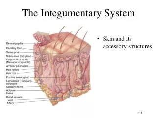
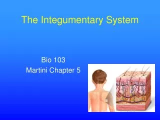
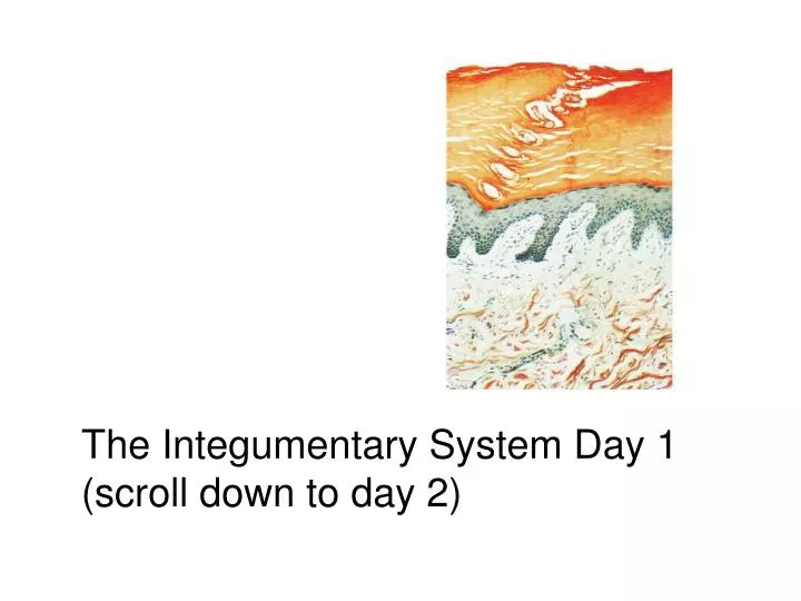
The Integumentary System Day 1 (scroll down to day 2). The Integumentary System. Integument is skin Skin and its appendages make up the integumentary system A fatty layer (hypodermis) lies deep to it Two distinct regions Epidermis Dermis. Functions of skin. Protection

E N D
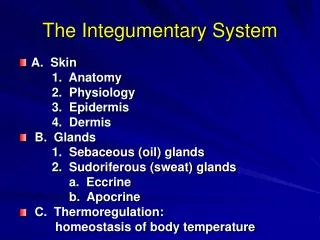
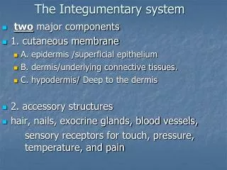
The Integumentary System • Integument is skin • Skin and its appendages make up the integumentary system • A fatty layer (hypodermis) lies deep to it • Two distinct regions • Epidermis • Dermis
Functions of skin • Protection • Cushions and insulates and is waterproof • Protects from chemicals, heat, cold, bacteria • Screens UV • Regulates body heat • Sensory reception (nerve endings) • Prevents unnecessary water loss and excretes small amounts of metabolic waste. • Synthesizes vitamin D with UV
Epidermis • Keratinized stratified squamous epithelium • Four types of cells • Keratinocytes – deepest, produce keratin (tough fibrous protein) • Melanocytes - make dark skin pigment melanin • Merkel cells – associated with sensory nerve endings • Langerhans cells – macrophage-like dendritic cells (see figure on next slide)
Epidermis • Layers (from deep to superficial) • Stratum basale (bas= latin for bottom/base) – single row of cells attached to dermis; youngest cells • Stratum spinosum (spin =latin for spiny) –mulitlayered cuboidal cells. Molecular bridges are spiny. Dark nucleipynknosisindication of cell death (see figure on next slide)
Epidermis • Layers (from deep to superficial) continued • Stratum granulosum(latin= small grains) – 3-5 layers of flattened keratinocytes producing keratin (hair and nails made of it also) • Stratum lucidum(latin= clear/transparent) -only on palms and soles. 3-5 layers of flat dead cells. • Stratum corneum – horny layer (cells dead, many layers thick, 20-50 rows). Sloughed off by normal wear and tear. (see figure on next slide)
Epidermis Continued • From stratum basale to slough in approximately 3 weeks. • Keratin prevents water loss • However the barrier is NOT absolute • Example: hot tub internal H20surfacewrinkle
Skin Color Primarily an inherited trait Secondarily regulated by the pituitary gland Melanocyte (greek=melano=dark) located in basal layer, produces melanin. Color of skin is determined by amount of melanin secreted NOT the number of melanocytes.
Yesterday you should have been able to answer… What are the five major functions of the integumentary system? Why do cells die as they are pushed farther from the stratum basale? What factors determine the color of your skin?
Today you will be able to answer… How is the dermis different from the epidermis? What role do blood vessels play in the dermis? How can you distinguish between the two areas of dermis? What product does a sebaceous gland secrete and what is its function? How may the two types of sweat glands be distinguished? What general function is performed by the skin receptors?
Remember… • Four basic types of tissue • Epithelium – epidermis just discussed • Connective tissue - dermis • Muscle tissue • Nervous tissue
Dermis • Strong, flexible connective tissue: your “hide” • Cells: fibroblasts, macrophages, mast cells, WBCs • Fiber types: collagen, elastic, reticular • Rich supply of nerves and vessels • Critical role in temperature regulation (the vessels) • Two layers (see next slides) • Papillary – areolar connective tissue; includes dermal papillae • Reticular – “reticulum” (network) of collagen and reticular fibers
*Dermal papillae *Dermis layers * *
Epidermis and dermis of (a) thick skin and (b) thin skin (which one makes the difference?)
Fingerprints, palmprints, footprints • Dermal papillae lie atop dermal ridges • Elevate the overlying epidermis into epidermal ridges • Are “sweat films” because of sweat pores • Genetically determined Flexion creases • Deep dermis, from continual folding Fibers • Collagen: strength and resilience • Elastic fibers: stretch-recoil • Striae: stretch marks • Tension lines (or lines of cleavage) • The direction the bundles of fibers are directed The dermis is the receptive site for the pigment of tattoos
Hypodermis • “Hypodermis” (Gk) = below the skin • “Subcutaneous” (Latin) = below the skin • Also called “superficial fascia” “fascia” (Latin) =band; in anatomy: sheet of connective tissue • Fatty tissue which stores fat and anchors skin (areolar tissue and adipose cells) • Different patterns of accumulation (male/female)
Skin appendages • Derived from epidermis but extend into dermis • Include • Hair and hair follicles • Sebaceous (oil) glands • Sweat (sudoiferous) glands • Nails
Nails • Of hard keratin • Corresponds to hooves and claws • Grows from nail matrix
Hair and hair follicles: complexDerived from epidermis and dermisEverywhere but palms, soles, nipples, parts of genitalia *“arrector pili” is smooth muscle * Hair bulb: epithelial cells surrounding papilla Hair papilla is connective tissue________________
Functions of hair • Warmth – less in man than other mammals • Sense light touch of the skin • Protection - scalp • Parts • Root imbedded in skin • Shaft projecting above skin surface • Make up of hair – hard keratin • Three concentric layers • Medulla (core) • Cortex (surrounds medulla) • Cuticle (single layers, overlapping)
Types of hair • Vellus: fine, short hairs • Intermediate hairs • Terminal: longer, courser hair • Hair growth: averages 2 mm/week • Active: growing • Resting phase then shed • Hair loss • Thinning – age related • Male pattern baldness • Hair color • Amount of melanin for black or brown; distinct form of melanin for red • White: decreased melanin and air bubbles in the medulla • Genetically determined though influenced by hormones and environment
Sebaceous (oil) glands • Entire body except palms and soles • Produce sebum by holocrine secretion • Oils and lubricates
Sweat glands • Entire skin surface except nipples and part of external genitalia • Prevent overheating • 500 cc to 12 l/day! (is mostly water) • Humans most efficient (only mammals have) • Produced in response to stress as well as heat
Types of sweat glands • Eccrine or merocrine • Most numerous • True sweat: 99% water, some salts, traces of waste • Open through pores • Apocrine • Axillary, anal and genital areas only • Ducts open into hair follices • The organic molecules in it decompose with time - odor • Modified apocrine glands • Ceruminous – secrete earwax • Mammary – secrete milk