Download
1 / 20
200 likes | 215 Vues
Learn about the historical aspects and definitions of contrast media in radiography, including subject contrast, contrast media properties, and different types of contrast media used in GI exams. Explore the use of negative and positive contrast mediums, and understand their properties and effects.

E N D
CONTRAST MEDIUM RAD 355
References • Radiographic procedures: By Stephen Chapman • Positioning in Radiography: By k.C.clarke. • Text book of radiographic positioning and related • anatomy; bykenneth L.Bontrager, John P. Lampignano, evolve, ELSEVIER MOSBY • 6th edition • Websites • http://www.e-radiography.net/
Learning objectives • By the end of these slides the student will be able to: • Know something about the historical aspects of contrast media • Define the term contrast • Differentiate between subject and radiographic contrast • Explain subject contrast chart • Differentiate between long scale and short scale contrast • Explain contrast Media Properties • Explain different types of contrast media used in GIT exams • Explain pharmacological agents used in GIT exams
Historical Aspects of Contrast Media • 1896 lead acetate was used as contrast medium which was toxic. • Walter Canon began to use bismuth subnitrate toxic. • By 1910 Barium sulphate began to appear as high atomic number, lack of toxicity, low cost, and its availability. • Walter Dandy 1918 use Air negative contrast medium. • Later, CO2, nitrous oxide, and oxygen came into use. • 1927 water soluble iodinated compounds were used but sodium iodide proved to be a blood vessel irritant.
Contrast • Definitions • "contrast" • exhibit noticeable differences when compared • "radiographic contrast" • visible differences between densities on an image • "subject contrast" • difference in the transmission of x rays due to the tissue type in the body part • “Contrast medium” • chemical compounds that permit visualization of the details of the internal structure or organs that would not otherwise be demonstrable.
Subject Contrast Definitions • Radiolucent (negative) • tissues that x rays easily penetrate • appear dark gray to black on the image • Radiopaque (positive) • tissues that x rays do not penetrate easily • appear light gray to white on the image • High atomic number
Tissue characteristics The body absorbs x-ray photons according to: 1.The various tissue atomic numbers. and 2. The amount of matter per volume of tissue. Higher atomic number tissues absorb more x-ray photons (e.g. bone) than low atomic number tissues (e.g. soft tissue) which transmit or scatter radiation. Radiographic images of anatomic areas classified as low in subject contrast result in few density differences and are difficult to visualize.
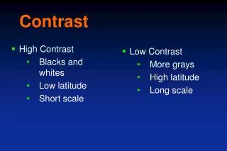
Radiation Qualitythe ability of a beam of radiographs to allow the production of diagnostically useful radiographs • Low kVp=less scatter=less penetration=high contrast • High contrast • Black and White • Short Scale • Great differences in adjacent structures • High kVp • Low contrast • Many shades of gray • Long Scale contrast: increased gradations of gray bet. Black and white on a radiographs. • Little differences in adjacent structures
Over Penetrated Too High kVp • The tissue types will determine how much kVp is needed. There are four basic tissue types. • Air filled least dense • Fat more dense • Muscle more dense • Bone most dense
kVp and Tissue Density • As we age, we loose bone and muscle mass, the kVp is reduced to compensate for this. • Very muscular patients require more kVp to assure proper penetration. • Disease processes that impact bone and tissue density will require adjustment of the kVp.
Radiographic Contrast vs. Contrast Media • Radiographic Contrast: Difference between adjacent densities in a radiograph. • The films or images have different levels of density – different shades of gray • X-rays show different features of the body in various shades of gray. • The gray is darkest in those areas that do not absorb X-rays well – and allow it to pass through • The images are lighter in dense areas (like bones) that absorb more of the X-rays.
Purpose of contrast media These diagnostic agents that are instilled into body orifices or injected into the vascular system, joints, and ducts to enhance subject contrast in anatomic areas where low subject contrast exists. The ability of the contrast media to enhance subject contrast depends on: The concentration of atoms of the element per volume of the medium The atomic number of the element
Purpose of Contrast Media • To enhance subject contrast or render high subject contrast in a tissue that normally has low subject contrast.
With Contrast Without Contrast IV Injection
With Contrast Without Contrast
Contrast Media Properties • able to show organ better • physiologically • no permanent alteration of organ • non toxic • able to be eliminated / excreted
Contrast Media Negative contrast • Radiolucent -AIR • Low atomic # material • Black on film • E.g. Air, CO2 Positive contrast • Radiopaque -BARIUM • High atomic # material • White on film • BaSO4
Negative Contrast Medium Air Pump