Download
1 / 1
10 likes | 155 Vues
No. 021. Diffusion-Weighted Imaging in Magnetic Resonance Imaging for prostate gland in Malaysian males with high prostate specific antigen in the diagnosis of prostate cancer. Keng Lim Ng1, Kheng Guan Tan2, Muhammad Nazri2, Shaik Ismail Bux2, Azad Hassan Razack1.

E N D
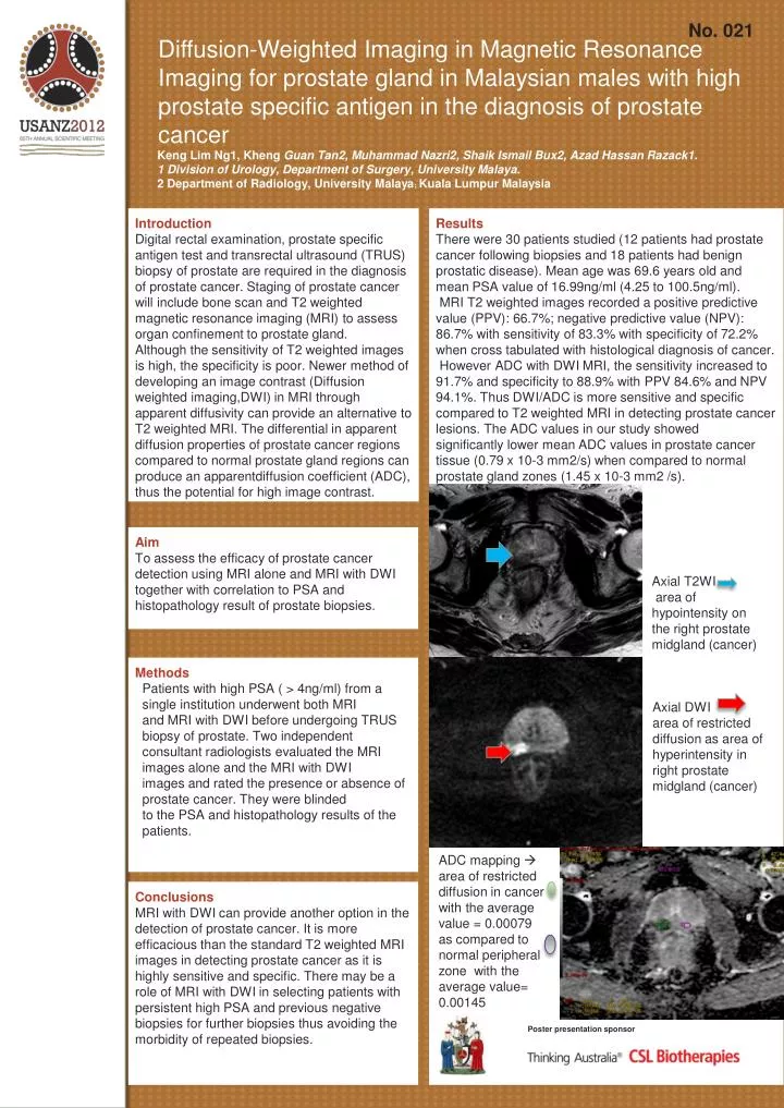
No. 021 Diffusion-Weighted Imaging in Magnetic Resonance Imaging for prostate gland in Malaysian males with high prostate specific antigen in the diagnosis of prostate cancer Keng Lim Ng1, KhengGuan Tan2, Muhammad Nazri2, Shaik Ismail Bux2, Azad Hassan Razack1. 1 Division of Urology, Department of Surgery, University Malaya. 2 Department of Radiology, University Malaya; Kuala Lumpur Malaysia Introduction Digital rectal examination, prostate specific antigen test and transrectal ultrasound (TRUS) biopsy of prostate are required in the diagnosis of prostate cancer. Staging of prostate cancer will include bone scan and T2 weighted magnetic resonance imaging (MRI) to assess organ confinement to prostate gland. Although the sensitivity of T2 weighted images is high, the specificity is poor. Newer method of developing an image contrast (Diffusion weighted imaging,DWI) in MRI through apparent diffusivity can provide an alternative to T2 weighted MRI. The differential in apparent diffusion properties of prostate cancer regions compared to normal prostate gland regions can produce an apparentdiffusion coefficient (ADC), thus the potential for high image contrast. Results There were 30 patients studied (12 patients had prostate cancer following biopsies and 18 patients had benign prostatic disease). Mean age was 69.6 years old and mean PSA value of 16.99ng/ml (4.25 to 100.5ng/ml). MRI T2 weighted images recorded a positive predictive value (PPV): 66.7%; negative predictive value (NPV): 86.7% with sensitivity of 83.3% with specificity of 72.2% when cross tabulated with histological diagnosis of cancer. However ADC with DWI MRI, the sensitivity increased to 91.7% and specificity to 88.9% with PPV 84.6% and NPV 94.1%. Thus DWI/ADC is more sensitive and specific compared to T2 weighted MRI in detecting prostate cancer lesions. The ADC values in our study showed significantly lower mean ADC values in prostate cancer tissue (0.79 x 10⁻³ mm2/s) when compared to normal prostate gland zones (1.45 x 10⁻³ mm2 /s). Aim To assess the efficacy of prostate cancer detection using MRI alone and MRI with DWI together with correlation to PSA and histopathology result of prostate biopsies. Axial T2WI area of hypointensity on the right prostate midgland (cancer) Methods Patients with high PSA ( > 4ng/ml) from a single institution underwent both MRI and MRI with DWI before undergoing TRUS biopsy of prostate. Two independent consultant radiologists evaluated the MRI images alone and the MRI with DWI images and rated the presence or absence of prostate cancer. They were blinded to the PSA and histopathology results of the patients. Axial DWI area of restricted diffusion as area of hyperintensity in right prostate midgland (cancer) ADC mapping area of restricted diffusion in cancer with the average value = 0.00079 as compared to normal peripheral zone with the average value= 0.00145 Conclusions MRI with DWI can provide another option in the detection of prostate cancer. It is more efficacious than the standard T2 weighted MRI images in detecting prostate cancer as it is highly sensitive and specific. There may be a role of MRI with DWI in selecting patients with persistent high PSA and previous negative biopsies for further biopsies thus avoiding the morbidity of repeated biopsies. Poster presentation sponsor