Download
1 / 45
500 likes | 1.02k Vues
Folding. Judith Klein-Seetharaman Department of Structural Biology jks33@pitt.edu. Objectives of this Lecture. Overview Folding/Misfolding Anfinsen Levinthal Paradox Folding Models The denatured state The molten globule Two-state folding Deciphering complex folding pathways.

E N D
Folding Judith Klein-SeetharamanDepartment of Structural Biology jks33@pitt.edu
Objectives of this Lecture • Overview Folding/Misfolding • Anfinsen • Levinthal Paradox • Folding Models • The denatured state • The molten globule • Two-state folding • Deciphering complex folding pathways Molecular Biophysics III – Klein-Seetharaman – Folding Lecture
Objectives of this Lecture • Overview Folding/Misfolding • Anfinsen • Levinthal Paradox • Folding Models • The denatured state • The molten globule • Two-state folding • Deciphering complex folding pathways Molecular Biophysics III – Klein-Seetharaman – Folding Lecture
Overview http://www-nmr.cabm.rutgers.edu/academics/biochem694/2006BioChem412/Biochem.412_2-24-2006lecture.pdf Molecular Biophysics III – Klein-Seetharaman – Folding Lecture
Objectives of this Lecture • Overview Folding/Misfolding • Anfinsen • Levinthal Paradox • Folding Models • The denatured state • The molten globule • Two-state folding • Deciphering complex folding pathways Molecular Biophysics III – Klein-Seetharaman – Folding Lecture
Oznur’s slide: Anfinsen’s Experiment Addition of mercaptoethanol and urea Removal of mercaptoethanol and urea Native, catalytically active state. Refolded correctly! Native, catalytically active ribonuclease A Unfolded; catalytically inactive. Reduced disulfide bonds. 1/105 random chance Folding is encoded in the amino acid sequence. Native state is the minimum energy state. Anfinsen, 1973. Molecular Biophysics III – Klein-Seetharaman – Folding Lecture
Objectives of this Lecture • Overview Folding/Misfolding • Anfinsen • Levinthal Paradox • Folding Models • The denatured state • The molten globule • Two-state folding • Deciphering complex folding pathways Molecular Biophysics III – Klein-Seetharaman – Folding Lecture
Oznur’s slide: How does a protein fold?Levinthal’s Paradox • Assume a chain of 100 amino acids. • Allow only 3 conformations. • - Possible conformations = 3100 ~ 1048 • Assume bond rotation rate 1014 sec. • - Reaching the native state would take: • 1026 years !Longer than the age of • the universe! Simplest case: random-walk Energy Entropy Protein folding cannot be random-walk. Dill & Chan, 1997 Levinthal, 1968 Molecular Biophysics III – Klein-Seetharaman – Folding Lecture
Objectives of this Lecture • Overview Folding/Misfolding • Anfinsen • Levinthal Paradox • Folding Models • The denatured state • The molten globule • Two-state folding • Deciphering complex folding pathways Molecular Biophysics III – Klein-Seetharaman – Folding Lecture
Oznur’s slide: The Three Protein Folding Models Framework model Hydrophobic collapse model Nucleation condensation model http://www.makro.ch.tum.de/users/BFHZ/Scheibel/Scheibel%202003%20Bordeaux-1.pdf Molecular Biophysics III – Klein-Seetharaman – Folding Lecture
Objectives of this Lecture • Overview Folding/Misfolding • Anfinsen • Levinthal Paradox • Folding Models • The denatured state • The molten globule • Two-state folding • Deciphering complex folding pathways Molecular Biophysics III – Klein-Seetharaman – Folding Lecture
Oznur’s slide: Random Coil and Denatured State Flory’s isolated pair hypothesis Rg values determined by SAXS “Φ,Ψ angles of each residue is sterically independent” There should not exist any non-local interactions. Rg values of 28 denatured proteins obeys the Flory’s power law. • Rg= RgNv • N = Length (Residues) • v = Solvent viscosity parameter Sosnick, T.R., et al. 2004 Flory, 1969. Molecular Biophysics III – Klein-Seetharaman – Folding Lecture
Oznur’s slide: Testing the random coil statistics For a protein ≈8% of the residues are varied; the remaining ≈92% of the residues remained fixed in their native conformation. 33 proteins Number of residues Simulated Rg follows the power law. Despite 92% of the native structure kept, random coil statistics are obtained. Fitzkee, N.C. and Rose, G.D. 2004 Molecular Biophysics III – Klein-Seetharaman – Folding Lecture
Oznur’s slide: The Denatured StateDoes Flory’s hypothesis hold? Conformations of polyalanine chains are enumerated to test the hypothesis. + ={A,G,M,R,L,F,E,K,Q} * = {J,P,O,I,o} Flory’s hypothesis is not valid for polypeptide chains. Backbone conformations are limited by additional steric clashes. Pappu et.al 2003. Molecular Biophysics III – Klein-Seetharaman – Folding Lecture
Which NMR spectrum is of folded and which is of unfolded lysozyme? Molecular Biophysics III – Klein-Seetharaman – Folding Lecture
Which NMR spectrum is of folded and which is of unfolded lysozyme? folded unfolded Molecular Biophysics III – Klein-Seetharaman – Folding Lecture
How would you use NMR to test for residual structure? Molecular Biophysics III – Klein-Seetharaman – Folding Lecture
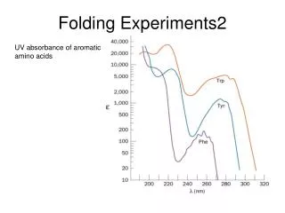
How would you identify residual structure in unfolded proteins with NMR? • What types of NMR parameters do you know? • chemical shifts • coupling constants • HetNOE • longitudinal relaxation rates (R1) • transverse relaxation rates (R2) Molecular Biophysics III – Klein-Seetharaman – Folding Lecture
How would you identify residual structure in unfolded proteins with NMR? • 1. Measurement of NMR parameters in 15N-labeled unfolded protein • chemical shifts • coupling constants • HetNOE • longitudinal relaxation rates (R1) • transverse relaxation rates (R2) • 2. Comparison of NMR parameters with random coil • 3. Deviation from random coil identifies residual structure • Application to unfolded conformations of hen egg white lysozyme: • oxidized in 8M urea • reduced and methylated in 8M urea • reduced and methylated in water Molecular Biophysics III – Klein-Seetharaman – Folding Lecture
Chemical shift differences between unfolded lysozyme and random coil Molecular Biophysics III – Klein-Seetharaman – Folding Lecture
Dynamics in folded/unfolded lysozyme Unfolded: Arrows indicate oxidized (all disulfide bonds present) lysozyme Folded: Molecular Biophysics III – Klein-Seetharaman – Folding Lecture
Relaxation Rates in Unfolded Lysozyme Unfolded lysozyme can be studied in 8 M urea. Unfolded lysozyme can also be studied without urea, if the disulfide bonds are reduced and the cysteines are derivatized to prevent them from forming disulfide bonds. Molecular Biophysics III – Klein-Seetharaman – Folding Lecture
Relaxation Rates in Unfolded Lysozyme What do you observe? Molecular Biophysics III – Klein-Seetharaman – Folding Lecture
Relaxation Rates in Unfolded Lysozyme Regions with higher relaxation rates are localized as clusters. Presence of clusters of residual structure that are restricted in conformational space, thus relax faster. Molecular Biophysics III – Klein-Seetharaman – Folding Lecture
How would you analyze the relaxation data? Molecular Biophysics III – Klein-Seetharaman – Folding Lecture
What are the assumptions of the model-free approach? Molecular Biophysics III – Klein-Seetharaman – Folding Lecture
Analysis of the relaxation data Three means of analysis have been proposed: • Model-free approach • Cole-Cole distributions • Gaussian clusters However: What gives rise to these clusters is not known. Molecular Biophysics III – Klein-Seetharaman – Folding Lecture
3. 2. 5. 4. 6. 1. Random Coil Model of Segmental Motion + Gaussian Distributions of Deviations 2 - - | i x | | i j | N 0 - - å å = + l R ( i ) R e Ae b int rinsic = j 1 x 0 Relaxation Rates in Unfolded Lysozyme There are six clusters of residual structure in HEWL-SME. Molecular Biophysics III – Klein-Seetharaman – Folding Lecture
Mapping of residual structure on the native structure Molecular Biophysics III – Klein-Seetharaman – Folding Lecture
Hydrophobic clusters of residual structure Molecular Biophysics III – Klein-Seetharaman – Folding Lecture
What stabilizes the clusters of residual structure? Molecular Biophysics III – Klein-Seetharaman – Folding Lecture
What stabilizes the clusters of residual structure? • Long-range interactions? • Local structure? • How would you test this? Molecular Biophysics III – Klein-Seetharaman – Folding Lecture
Approach 1 • Peptides: if peptides without structural context of the full chain contain structure, then this structure is independent of long-range stabilization Molecular Biophysics III – Klein-Seetharaman – Folding Lecture
Approach 2 • Test for the presence of long-range interactions in the context of the full-length protein • What approaches can you imagine to test for long-range interactions? Molecular Biophysics III – Klein-Seetharaman – Folding Lecture
Residual Structure Mapped onto Native Structure Clusters of deviations from random coil dynamics map onto proximal regions in the native structure, except cluster 3. Molecular Biophysics III – Klein-Seetharaman – Folding Lecture
How would you test for the presence of long-range interactions?Approach 1. Study effect of mutation Molecular Biophysics III – Klein-Seetharaman – Folding Lecture
Effect of mutation on chemical shifts Molecular Biophysics III – Klein-Seetharaman – Folding Lecture
Effect of mutation on relaxation rates A single point mutation, W62G in cluster 3, disrupts all clusters in reduced and methylated lysozyme. Molecular Biophysics III – Klein-Seetharaman – Folding Lecture
Effect of mutation on chemical shifts Molecular Biophysics III – Klein-Seetharaman – Folding Lecture
Effect of mutation on relaxation rates Molecular Biophysics III – Klein-Seetharaman – Folding Lecture
Model for unfolded ensemble Molecular Biophysics III – Klein-Seetharaman – Folding Lecture
Compactness by NMR Molecular Biophysics III – Klein-Seetharaman – Folding Lecture
Approach 2. FRET - So far has been only used for global changes, not to detect specific contact formation Haustein and Schwille (2004) Current Opin. Structural Biology 14, 531-540. Molecular Biophysics III – Klein-Seetharaman – Folding Lecture
Approach 3. EPR – proton relaxation interaction up to 20-25Å Staphylococcus nuclease – Gillespie and Shortle (1997) JMB 268, 170-184 and 158-169. Molecular Biophysics III – Klein-Seetharaman – Folding Lecture
A. Wild-Type B. W62G Role of disulfide bonds for dynamics Disulfide bonds and hydrophobic clusters are cooperative. Molecular Biophysics III – Klein-Seetharaman – Folding Lecture