Download
1 / 29
290 likes | 544 Vues
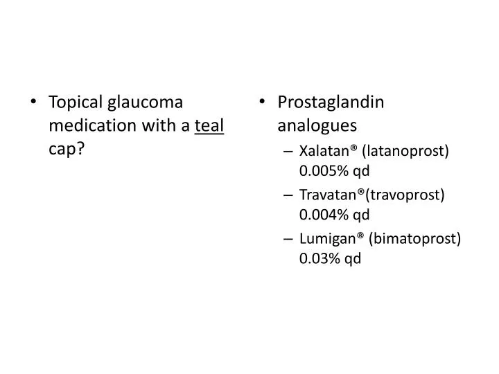
Topical glaucoma medication with a teal cap?. Prostaglandin analogues Xalatan ® ( latanoprost ) 0.005% qd Travatan®(travoprost ) 0.004% qd Lumigan ® ( bimatoprost ) 0.03% qd. Mechanism of action: Prostaglandin analogues. Enhances uveoscleral outflow.

E N D
Topical glaucoma medication with a teal cap? • Prostaglandin analogues • Xalatan® (latanoprost) 0.005% qd • Travatan®(travoprost) 0.004% qd • Lumigan® (bimatoprost) 0.03% qd
Mechanism of action: Prostaglandin analogues • Enhances uveoscleral outflow.
Topical glaucoma medication with a yellowcap? • non-selective beta blockers 0.50% • Timoptic® (timolol) • Betagan® (levobunolol) • β2 selective beta blockers 0.50% • Betoptic S® (betaxolol)
Topical glaucoma medication with a blue cap? • non-selective beta blockers 0.25% • Timoptic® (timolol) • Betagan® (levobunolol) • β2 selective beta blockers 0.25% • Betoptic S® (betaxolol)
Mechanism of action of beta blockers? • reduces aqueous production (by blocking beta-2 receptors on nonpigmentedciliary epithelium)
Topical glaucoma medication with a purple cap? • α2 adrenergic agonists • Iopidine® (apraclonidine) • 0.5% tid • 1.0% bid • Alphagan-P® (brimonidine) • 0.1% • 0.15% • 0.2% • bid or tid
Mechanism of action for α2 adrenergic agonists • decreases aqueous production • increases uveoscleral outflow • “Both kinds of glaucoma treatment!”
topical glaucoma medication with a green cap? • Cholinergic agonists (miotics) • pilocarpine (0.25% -- 10%) 2% or 4% most often. • carbachol • echothiophate
Mechanism of action for cholinergic agonists • increases trabecular outflow • mayincreasuveoscleral outflow
Topical glaucoma medication with an orange cap? • carbonic anhydrase inhibitors (CAIs) • Trusopt® (dorzolamide) 2% • Azopt® (brinzolamide) 1%
Mechanism of action for carbonic anhydrase inhibitors • decreases aqueous production
Glaucoma medications that increase outflow • Uveoscleral: • prostaglandins • α2 adrenergic agonists • Trabecular: • cholinergic agonists / miotics
Glaucoma medications that reduce aqueous production • β blockers • α2 adrenergic agonists • carbonic anhydrase inhibitors
“Discussion” points for every glaucoma patient: • “controlling a risk factor (high eye pressure) for glaucoma” • “in hopes to prevent vision loss” • “no guarantee that vision loss will not occur” • “like controlling (blood pressure) a risk factor for heart attack and stroke”
Glaucoma standard of care • dilation with retinal biomiscropy • Goldmanntonometry • visual fields • gonioscopy • pachymetry • photography • (debatable) OCT/GDx/HRT
OHTS goals of study • Ocular Hypertension Treatment Study • Evaluate safety / efficacy of topical glaucoma medications • to prevent or delay congenital open angle glaucoma (COAG) in patients with high IOP • identify baseline demographic and clinical factors • predict which patients will develop COAG
Ocular Hypertension Treatment Study (OHTS) • OHTS Enrollees • patients with: • high IOP • normal visual fields (VF) • normal discs • patient randomly assigned to medical treatment or observation • patient monitored: • VF q 6 months • fundus photos q year • endpoint of COAG: • VF evidence • disc evidence • or both VF and disc evidence
Ocular Hypertension Treatment Study (OHTS) • OHTS results • COAG patients: • 55% diagnosed via disc changes without VF changes • 20% reduction in IOP decreased incidence of COAG by 50% at 5 years • black people developed COAG at significantly higher rate
Ocular Hypertension Treatment Study (OHTS) • OHTS Important risk factors for glacomatous damage in ocular hypertensives • higher IOP • old age • large C/D • greater pattern standard deviation • thin central cornea thickness (CCT)
Ocular Hypertension Treatment Study (OHTS) • OHTS central corneal thickness (CCT) findings • patients with CCT less than 555 are 3x more likely to develop POAG • Much bigger risk factor than: • race • family history • refractive error • general health • Recommendation: pachymetry as standard part of evaluation of ocular hypertensive patients.
Ocular Hypertension Treatment Study (OHTS) • OHTS Guidelines • IOP: • 23.75 or below low risk • 23.75 < IOP < 25.75 moderate risk • above 25.75 high risk • CCT (pachymetry): • above 588 low risk • 555 < CCT < 588 moderate risk • 555 or below high risk • vertical C/D • 0.3 or less low risk • 0.3 < C/D < 0.5 moderate risk • 0.5 or above high risk
In glaucoma, NFL damage precedes… • field loss and disc changes • 5 year, 50% “rule” 50% of NFL is lost before a VF will reveal a defect. • “we are in the business of preventing any vision loss”
Instruments commonly used to manage patients with glaucoma • HRT2 – Heidelbert Retinal Tomograph • Stratus OCT – Optical Coherence Tomography from Zeiss Humphrey • GDx-VCC nerve fiber analyzer (with variable corneal compensator from Laser Diagnostic Technologies)
HRT, OCT, GDx factoids • image the optic disc and/or retinal NFL • each has strengths & weaknesses • useful for glaucoma suspects moderate glaucoma patients • NOT helpful for advanced glaucoma patients • cost: HRT < GDx < OCT
Optic nerve imaging considerations • imaging reimbursable at least once a year, HOWEVER, cannot be reimbursed for fundus photos & imaging on same day. • results need to be used within a larger clinical context! • ONH tissue, VF defects and imaging should all correlate & “make sense”
HRT strengths & “considerations” • The HRT utilizes confocal scanning laser ophthalmoscopy • Strengths • shows topography of optic nerve • detects patient progression • confirms nerve size • evaluates cupping • “Considerations” • accuracy depends on drawing contour line correctly the first time. • probably not the best for assessing thickness of RNFL
OCT strengths & “considerations” • OCT uses optical back scattering of light to compute tomographic images based on amount of incident light reflected by tissue. • Strengths • accurately measures RNFL thickness • correlates highly with VF loss & disc damage • reproducible • image quality monitored during test • awesome for macular holes, edema CNV mgmt • reason why less fewer FLANs done at UMSL • “Considerations” • automatically generated disc contour lies can be somewhat inaccurate • does not determine progression • difficult to determine how much change is clinically significant • requires dilated pupil, minimal media opacity
GDx strengths & “considerations” • GDx uses a scanning laser polarimeter • Strengths • measures RNFL • very reliable • portable • can do undilated • media opacities least likely to interfere • Considerations • image quality cannot be measured during test
GDx NFL analysis • Gives ‘NFI’ number: • 0—30 normal, low probability of glaucoma • 31—70 glaucoma suspect • 71—100 high probability of glaucoma • The NFI number does NOT indicate severity or progression of glaucoma.