Download
1 / 34
340 likes | 477 Vues
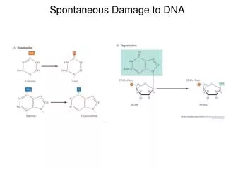
Oxidative Damage of DNA. Oxidative damage results from aerobic metabolism, environmental toxins, activated macrophages, and signaling molecules (NO). Compartmentation limits oxidative DNA damage. Oxidation of Guanine Forms 8-Oxoguanine. The most common mutagenic base lesion is 8-oxoguanine.

E N D
Oxidative Damage of DNA Oxidative damage results from aerobic metabolism, environmental toxins, activated macrophages, and signaling molecules (NO) Compartmentation limits oxidative DNA damage
Oxidation of Guanine Forms 8-Oxoguanine The most common mutagenic base lesion is 8-oxoguanine guanine 8-oxoguanine from Banerjee et al., Nature 434, 612 (2005)
Repair of 8-oxo-G Replication of the 8-oxoG strand preferentially mispairs with A and mimics a normal base pair and results in a G-to-T transversion 8-oxoguanine DNA glycosylase/ b-lyase (OGG1) removes 8-oxo-G and creates an AP site MUTYH removes the A opposite 8-oxoG from David et al., Nature447, 941 (2007)
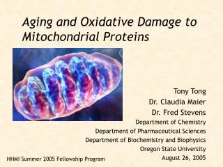
MTH1 Prevents Incorporation of Oxidized dNTPs into DNA Free dNTPs are much more susceptible to oxidative damage than bases in duplex DNA Oxidized precursors are misincorporated and are mutagenic from Dominissini and He, Nature508, 191 (2014) MTH1 removes oxidized nucleotides from the pool
Inhibition of MTH1 Selectively Kills Cancer Cells MTH1 is not essential in normal cells Higher levels of ROS in cancer cells causes a non-oncogene addiction to MTH1 from Gad et al., Nature 508, 215 (2014)
UV-Irradiation Causes Formation of Thymine Dimers from Lodish et al., Molecular Cell Biology, 6th ed. Fig 4-38
Nonenzymatic Methylation of DNA Formation of 600 3-me-A residues/cell/day are caused by S-adenosylmethionine 3-me-A is cytotoxic and is repaired by 3-me-A-DNA glycosylase 7-me-G is the main aberrant base present in DNA and is repaired by nonenzymatic cleavage of the glycosyl bond
Effect of Chemical Mutagens Nitrous acid causes deamination of C to U and A to HX U base pairs with A HX base pairs with C
Repair Pathways for Altered DNA Bases from Lindahl and Wood, Science 286, 1897 (1999)
Direct Repair of DNA Photoreactivation of pyrimidine dimers by photolyase restores the original DNA structure O6-methylguanine is repaired by removal of methyl group by MGMT 1-methyladenine and 3-methylcytosine are repaired by oxidative demethylation
Base Excision Repair of a G-T Mismatch BER works primarily on modifications caused by endogenous agents At least 8 DNA glcosylases are present in mammalian cells DNA glycosylases remove mismatched or abnormal bases AP endonuclease cleaves 5’ to AP site AP lyase cleaves 3’ to AP site from Lodish et al., Molecular Cell Biology, 6th ed. Fig 4-36
DNA Glycosylases from Xu et al., Mech.Ageing Dev. 129, 366 (2008) Each glycosylase has limited substrate specificity, but there is redundancy in damage recognition
Mechanism of hOGG1 Action from David, Nature434, 569 (2005) hOGG1 binds nonspecifically to DNA Contacts with C results in the extrusion of corresponding base in the opposite strand G is extruded into the G-specific pocket, but is denied access to the oxoG pocket oxoG moves out of the G-specific pocket, enters the oxoG-specific pocket, and excised from the DNA
Nucleotide Excision Repair Repairs helix-distorting lesions UV-induced pyrimidine dimers Bulky adducts Intrastrand crosslinks ROS-generated cyclopurines
Global Genome NER – Damage Recognition Probes for helix distorting lesions XPC is the damage sensor which finds the ssDNA gap caused by disrupted pairing UV-DDB (DDB1 and DDB2) can stimulate XPC binding by extruding the lesion to create ssDNA from Marteijn et al., Nature Rev.Mol.Cell Biol. 15, 465 (2014)
Transcription-coupled NER – Damage Recognition Repairs transcription-blocking lesions CSB, UVSSA and USP7 interact with Pol II With CSA, promotes backtracking of Pol II to expose lesion from Marteijn et al., Nature Rev.Mol.Cell Biol. 15, 465 (2014)
NER – Lesion Verification TFIIH complex is recruited to the lesion XPB and XPD are helicases with opposite polarity XPD verifies the existence of lesions and XPA binds to altered nucleotides RPA protects the undamaged strand from nucleases XPG nuclease binds to the complex from Marteijn et al., Nature Rev.Mol.Cell Biol. 15, 465 (2014)
NER – Strand Excision XPF nuclease is recruited by XPA and directed to the damaged strand by RPA XPF and XPG excises the lesion from Marteijn et al., Nature Rev.Mol.Cell Biol. 15, 465 (2014)
NER – Gap Filling and Ligation PCNA recruits DNA polymerase to fill ss gap Nick is sealed by DNA ligase from Marteijn et al., Nature Rev.Mol.Cell Biol. 15, 465 (2014)
Chromatin Dynamics in GG-NER NER is stimulated by an open chromatin environment UV-DDB ubiquitylates core histones and associates with PARP1 which PARylates chromatin Histone acetylation stimulates NER Chromatin remodelling complexes displace nucleosomes from Marteijn et al., Nature Rev.Mol.Cell Biol. 15, 465 (2014)
Clinical Implications of Defective NER GG-NER is elevated in germ cells to maintain the entire genome to prevent mutagenesis Defective GG-NER increases cancer predisposition Xeroderma pigmentosum TC-NER is elevated in somatic cells to repair expressed genes to prevent cell death Defective TC-NER causes premature cell death, neurodegeration and accelerates aging Cockayne Syndrome
Mismatch Repair Repairs DNA replication errors and insertion-deletion loops Decreases mutation frequency by 102 - 103 Plays a role in triplet repeat expansion, somatic hypermutation and class switch recombination
Mismatch repair in E. coli GATC sequences are methylated by dam methylase Newly replicated DNA is transiently hemimethylated MutS recognizes a mismatch or small IDL MutS bends DNA, recruits MutL and forms a small dsDNA loop MutH nicks the unmethylated GATC Helicase unwinds the nicked DNA which is degraded past the mismatch Gap is repaired by Pol III and ligase from Marra and Schar, Biochem.J. 338, 1 (1998)
Mismatch Repair in Eukaryotes MutS homologs recognize mismatch and form a ternary complex with MulL homologs and the mismatch PMS2 is a mismatch-activated strand- specific nuclease, and the break is directed to the strand contain the preexisting nick EXO1 excises the mismatch The gap is filled in by PCNA, Pold and DNA ligase Defective mismatch repair is the primary cause of certain types of human cancers from Hsieh and Yamane, Mech.Ageing Dev. 129, 391 (2008)
Causes of and Responses to ds Breaks DSBs result from exogenous insults or normal cellular processes DSBs result in cell cycle arrest, cell death, or repair Repair of DSBs is by homologous recombination or nonhomologous end joining from van Gent et al., Nature Rev.Genet. 2, 196 (2001)
Initiation of Double-stranded Break Repair MRN complex recognizes DSB ends and recruits ATM ATM phosphorylates H2A.X and recruits MDC1 to spread gH2A.X TIP60 and UBC13 modify H2A.X MDC1 recruits RNF8 which ubiquitylates H2A.X RNF168 forms ubiquitin conjugates and recruits BRCA1 from van Attikum and Gasser, Trends Cell Biol. 19, 204 (2009)
ATM Mediates the Cell’s Response to DSBs DSBs activate ATM ATM phosphorylation of p53, NBS1 and H2A.X influence cell cycle progression and DNA repatr from van Gent et al., Nature Rev.Genet. 2, 196 (2001)
Repair of ds Breaks by Homologous Recombination ssDNAs with 3’ends are formed and coated with Rad51, the RecA homolog Rad51-coated ssDNA invades the homologous dsDNA in the sister chromatid The 3’-end is elongated by DNA polymerase, and base pairs with ss 3-end of the other broken DNA DNA polymerase and DNA ligase fills in gaps from Lodish et al., Molecular Cell Biology, 5th ed. Fig 23-31
Role of BRCA2 in Double-stranded Break Repair BRCA2 mediates binding of RAD51 to ssDNA RAD51-ssDNA filaments mediate invasion of ssDNA to homologous dsDNA from Zou, Nature 467, 667 (2010)
Repair of ds Breaks by Nonhomologous End Joining KU heterodimer recognizes DSBs and recruits DNA-PK Mre11 complex tethers ends together and processes DNA ends DNA ligase IV and XRCC4 ligates DNA ends from van Gent et al., Nature Rev.Genet. 2, 196 (2001)
Translesion DNA Synthesis Replicative polymerase encounters DNA damage on template strand Catalytic site of replicative polymerases is intolerant of misalignment between template and incoming nucleotide Replicative polymerase is replaced by TLS polymerase which inserts a base opposite lesion Base pairing is restored beyond the lesion and replicative polymerase replaces TLS polymerase TLS can occur in S or G2 from Sale et al., Nature Rev.Mol.Cell Biol. 13, 141 (2012)
There are Multiple TLS Polymerases TLS polymerases are recruited by interactions with the sliding clamp There are multiple TLS polymerases TLS polymerases have low processivity and low fidelity, and lack 3’-5’ exonucleases TLS polymerases are selective for certain lesions from Sale et al., Nature Rev.Mol.Cell Biol. 13, 141 (2012) Most mutations caused by DNA lesions are caused by TLS polymerases
TLS Polymerases Can Be Accurate or Error-prone Pol k bypasses an abasic site and often causes a -1 frameshift Pol h bypasses a thymine dimer and inserts AA Pol i is accurate with dA template and error-prone with dT template Replicative polymerases insert dC or dA opposite 8-oxo-G, Pol i inserts dC The likelihood that TLS polymerases are error-prone depends on the nature of the lesion and the TLS polymerase that is utilized
Somatic Hypermutation of Ig Genes Depends on TLS Polymerases AID deaminates dC to dU Uracil DNA glycosylase forms an abasic site, and REV1 incorporates dC opposite the site MMR proteins lead to the formation of a ss gap, PCNA is ubiquitylated, and Pol h is recruited, generating mutations at A-T from Sale et al., Nature Rev.Mol.Cell Biol. 13, 141 (2012)