Download
1 / 40
400 likes | 411 Vues
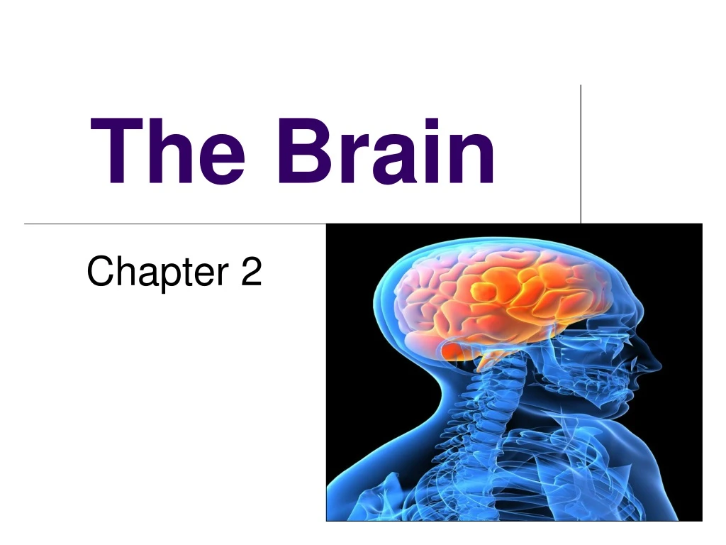
The Brain. Chapter 2. The Brain. Techniques for Studying the Brain. Methods. Brain research can be done in a variety of ways. Brain damage as a result of an accident or disease can provide a wealth of information. Lesioning is the removal or destruction of part of the brain .

E N D
The Brain Chapter 2
The Brain Techniques for Studying the Brain
Methods Brain research can be done in a variety of ways. Brain damage as a result of an accident or disease can provide a wealth of information. Lesioning is the removal or destruction of part of the brain. Any time brain tissue is removed (tumor, lobotomy, behavior experiment in animals, etc.) researchers can examine behavior changes and infer the function of that part of the brain.
Functional Methods EEG EEG (electroencephalogram) is an amplified recording of the electrical waves sweeping across the brain’s surface, measured by electrodes placed on the scalp (sleep studies, etc.)
PET Scan Functional Methods PET (positron emission tomography) Scan is a visual display of brain activity that detects a radioactive form of glucose while the brain performs a given task. By doing this, one can connect brain activity to the area of the brain that controls it.
MRI Scan Structural Methods MRI (magnetic resonance imaging) uses magnetic fields and radio waves to produce computer-generated images that distinguish among different types of brain tissue. Uses different technology to produce picture, but is similar to a CAT (computerized axial tomography). The first images is of a normal brain. The second image shows ventricular enlargement in a schizophrenic patient.
Combination Method (structure & function) fMRI An fMRI (functional MRI) is a comparison of shots before and during the performance of mental functions to map the parts of the brain that control those functions.It combines elements of the MRI (structure) and PET (function). The fMRI image shows brain regions that are active when a participants lies.
The Brain Areas and Parts of the Brain
I. Older Brain Structures A. The Brainstem 1. Medulla 2. Pons 3. Reticular Formation B. Thalamus C. Cerebellum D. The Limbic System 1. Amygdala 2. Hypothalamus 3. Hippocampus
The Brainstem TheBrainstemisthe oldest part of the brain, beginning where the spinal cord swells and enters the skull. It is responsible for automatic survival functions. brainstem http://www.npr.org/templates/story/story.php?storyId=129027124&sc=fb&cc=fp
Parts of the Brain Stem: The Medullais the base of the brainstem that controls heartbeat and breathing. Pons deals with facial expressions. Also plays a role in sleep & dreaming. Reticular Formationisa nerve network in the brainstem that plays an important role in controlling arousal. Pons
Parts of the Brain Stem The Thalamusis the brain’s sensory switchboard, located on top of the brainstem. It directs messages to the sensory areas in the cortex and transmits replies to the cerebellum and medulla. It receives information for all of the senses EXCEPT for smell.
Cerebellum The Cerebellum is called the “little brain” and is attached to the rear of the brainstem. It helps coordinate voluntary movements and balance. It also plays a part in memory, emotion regulation, timing, emotional modulation and sensory discrimination. Brainstem
The Limbic System The Limbic Systemisa doughnut-shaped system of neural structures at the border of the brainstem and cerebrum, associated with emotions such as fear, aggression and drives for food and sex. It includes thehippocampus, amygdala, andhypothalamus.
Hippocampus Hippocampus The Hippocampus processes memories.
Amygdala The Amygdalaconsists of two almond-shaped neural clusters linked to the emotions; particularary those of fear and anger.
Hypothalamus The Hypothalamus lies below (hypo) the thalamus. It directs several maintenance activities like eating, drinking, body temperature, and control of sexual arousal. It helps control the endocrine system by giving directions to the pituitary gland. Pituitary
The Limbic System contains many Reward/Pleasure Centers Olds and Milner (1954) discovered that Rats cross an electrified grid for self-stimulation when electrodes are placed in the reward (hypothalamus) center. When the limbic system is manipulated, a rat will navigate fields or climb up a tree (bottom picture). It is possible that some addictive behavior may be related to a genetic disorder (reward deficiency syndrome).
Cerebral Cortex The intricate fabric of interconnected neural cells that covers the cerebral hemispheres. It is the body’s ultimate control and information processing center.
Need More Mnemonics? • Cerebral Cortex: imagine a Texas cowboy hat on top of a brain. The cortex is the outer layer of the brain just under the hat where complex thinking occurs. • Corpus Callosum: The corpus callosum is the fibers that connect the two halves of the brain. Thus, it adds the two parts together. Think of the corPLUS CalloSUM. Since the corpus callosum coordinates communication between the two hemispheres, think of corpus Call Someone. • Thalamus: the thalamus takes sensations that come from the body and directs them to the appropriate part of the brain for processing. Thus, think of Hal and Amos – two traffic cops in the brain who direct these sensations to the right route. • Hypothalamus: the hypothalamus regulates a number of things in the body such as body temperature, thirst, hunger, and sex drive. Think of “hypo the llamas”. Your llamas are hot, sweaty and thirsty and you use a hypo to spray water on them to cool them down and give them some water. • Hippocampus: the hippocampus is the seat of memory. Think of a hippo with a compass. The hippo uses the compass to find his way back to the swamp because he can’t remember where it is. • Amygdala: the amygdala controls your sense of fear. Think of either a MIG coming right at you and, of course, making you afraid, or picture a scary wig with dollars in it • Pons: the pons helps you relax and sleep. Think of a relaxing pond. • Cerebellum: the cerebellum helps in coordination and balance. Picture your favorite athlete with bells all over his/her body (hanging from his/her clothes, hands, feet, etc.). • Reticular Formation: the reticular formation helps you to become alert and aroused when you need to be. Think of what would happen if you were napping and someone tickled you: your reticular formation would kick into gear to wake you up. • Medulla: the medulla regulates the autonomic activity of your heart and lungs. Picture medals over your heart and lungs, or stick those medals into a heart.
Structure of the Cerebral Cortex Each brain hemisphere is divided into four lobes that are separated by prominent fissures. These lobes are the: a. frontal lobe – judgement/reasoning b. parietal lobe – senses c. occipital lobe – vision d. temporal lobe – hearing A. B. C. D.
Functions of the Cerebral Cortex TheMotor Cortexis the area at the rear of the frontal lobes that control voluntary movements. The Sensory Cortex is the area at the front of the parietal lobes that receives information from skin surface and sense organs.
Functions of the Cerebral Cortex The visual cortex is located in the occipital lobe of the brain. The functional MRI scan shows the visual cortex is active as the subject looks at faces.
Functions of the Cerebral Cortex The auditory cortex is located in the temporal lobe of the brain. http://www.ted.com/talks/lang/eng/oliver_sacks_what_hallucination_reveals_about_our_minds.html
Association Areas The association areas integrate sensory information and stored memories. More intelligent animals have increased “uncommitted” or association areas of the cortex.
The Curious Story of Phineas Gage (1848) Frontal lobe damage showed effects on personality and social functioning
http://www.accessatlanta.com/celebrities-tv/brazilian-worker-survives-iron-1501218.htmlhttp://www.accessatlanta.com/celebrities-tv/brazilian-worker-survives-iron-1501218.html http://www.boston.com/news/health/articles/2009/07/22/newly_discovered_image_offers_fresh_insights_about_1848_medical_miracle/
Language Aphasiais an impairment of language, usually caused by left hemisphere damage either to Broca’s area (impaired speaking) or to Wernicke’s area (impaired understanding).
Specialization & Integration Brain activity when hearing, seeing, and speaking words.
The Brain’s Plasticity • The brain is sculpted by our genes but also by our experiences. • Plasticityrefers to the brain’s ability to modify itself after some type of injury or illness. • Usually the brain areas that are related to the damaged/missing part develop the ability to function as a part of the new system. For example, in blind people the visual cortex may register and process touch and/or hearing also (heightening those senses) • Our brains demonstrate more plasticity when we are children.
The Brain The Divided Brain
Our Divided Brain • Our brain is divided into two hemispheres. • The Left Hemisphere • Processes logical tasks (reading, writing, speaking, mathematics, and comprehension skills) • Controls the right side of our body • In the 1960s, it was termed as the dominant brain. • The Right Hemisphere • Processes non-verbal tasks/perceptual (spatial relationships, musical/artistic ability and mental imagery) • Controls the left side of our body • May also be related to some negative emotions • The Corpus Callosum is a wide band of axon fibers that connect the two hemispheres and allow them to communicate.
Splitting the Brain A procedure in which the two hemispheres of the brain are isolated by cutting the connecting fibers (mainly those of the corpus callosum) between them. Usually done to prevent uncontrollable seizures in patients with severe epilepsy. Corpus Callosum
6 Pop Quiz 7 5 4 3 2 1
Split Brain Patients With the corpus callosum severed, objects (apple) presented in the right visual field can be named. Objects (pencil) in the left visual field cannot.
Lateralization also Occurs in Non-Split Brains People with intact brains also show left-right hemispheric differences in mental abilities. A number of brain scan studies show normal individuals engage their right brain when completing a perceptual task and their left brain when carrying out a linguistic task.