Download
1 / 63
640 likes | 667 Vues
Check a few updates about the covid19 diagnosis by Dr. gurbilas. He is a famous gastroenterologist in Chandigarh.

E N D
Current updates on COVID-19 Diagnosis Dr.Gurbilas P. Singh, FRCP (London) Convener, GI Rendezvous Director Education & Training, GI, Liver specialist and Interventional Endoscopist, SarvhitGastrocity, Amritsar, India
Our experts – Special thanks Ms. Harvinder Kaur L.S. Malaysian Institute of Medical Laboratory Sciences, Malaysia Dr.Kanwardeep Singh, Professor of Microbiology, Pl, Viral Research and Diagnostic Lab. Government Medical College, Amritsar, India
COVID 19, A pandemic (Greek - pan "all" and demos - "people“) • A pandemic is the worldwide spread of a new disease as per WHO • The US Centers for Disease Control and Prevention defines a pandemic as “an epidemic that has spread over several countries or continents, usually affecting a large number of people.” • A widespread endemic disease with a stable number of infected people is not a pandemic.
COVID – 19 Diagnosis – what to expect ? • How to suspect and diagnose in clinical practice • Clinical manifestations and variations • Comorbidities and their impact on outcome • Current methods of surveillance
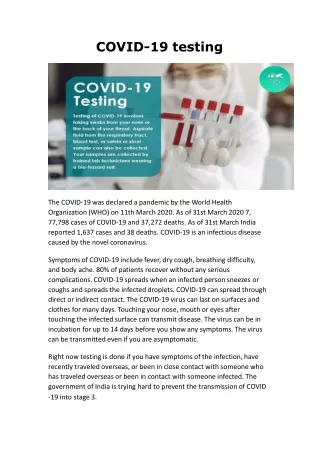
Background: COVID-19 • Newly emergent coronavirus, SARS-CoV-2 • Respiratory infection, including severe pneumonia • Respiratory droplets - sneezing, coughing, or talking • Spreads through touching a surface or object that has the virus on it and then touching own mouth, nose, or possibly eyes • Some individuals with coronavirus may be asymptomatic
HOW TO SUSPECT AND DIAGNOSE COVID IN DAILY PRACTICE • Common symptoms include cough, fever, chills, shortness of breath, muscle aches, sore throat, unexplained loss of taste or smell, diarrhea, and headache. • Symptoms can be mild and may aggravate over 5 to 7 days, sometimes worsening with pneumonia. • Approximately, 1 out of 5 infected individuals becomes seriously ill with difficulty in breathing, especially in the elderly with underlying health conditions REFRENCE: https://www.cdc.gov/coronavirus/2019-ncov/symptoms-testing/symptoms.html https://apps.who.int/iris/bitstream/handle/10665/331506/WHO-2019-nCoV-SurveillanceGuidance-2020.6-eng.pdf
Mr. B, 35 years male – Teleconsult • Symptoms of bloating • Heartburn • Nausea, especially on taking Paracetamol • Sore throat with occasional coughing • Dysphagia? – although only when he had to take medicines! • Background of low grade fever since 9 days ! • “But doctor it is not COVID. We got it checked and it is negative.”
TAKING History – No assumptions! • When did the fever start precisely? – “Saturday night” • When was COVID 19 test performed and what test was it? – “Tuesday AM and it was RT PCR test” • Any cough or breathlessness ? – “Not really, but I cough off and on during this season anyway, and oxygen levels are between 93% and 95%” • Family history – Both parents hypertensive and mom a diabetic. • Medical and personal history – Fit and well with no regular medication and no allergies. Non smoker and occasionally takes alcohol.
Management of Mr. B – COVID status A) Happy with the test done and we do not believe it is COVID 19. B) It could be COVID 19 because the saturations seem low! C) It is definitely COVID 19 because of the symptoms of cough and fever! D) It could be COVID 19 and the test may have been done too early!
Management of Mr. B – Next step? A) Repeat RT PCR for COVID 19 B) Do some routine bloods including CRP and D -Dimers to clinch the diagnosis. C) Do a chest X Ray as there are some respiratory symptoms and this would clinch the diagnosis. D) Do a CT Chest to assess the lung fields for any changes related to COVID 19.
Management of Mr. B – if Mr. B turns out to be COVID 19 positive, • Immaterial of what the clinical status is he should be admitted to a hospital as he is COVID positive. • Since there is strong family history of diabetes and hypertension and hence best to watch him in the hospital. • For his symptoms of dyspepsia, to keep a close watch on him he should be admitted to a hospital. • He should be given an emergency (helpline) number and advised to closely watch his oxygen levels, reporting any changes in his symptoms of cough and shortness of breath.
Clinical classifications Asymptomatic infection (silent infection) • Testing positive for SARS-CoV-2, but without clinical symptoms or abnormal chest imaging findings. Acute upper respiratory tract infection • With only fever, cough, pharyngeal pain, nasal congestion, fatigue, headache, myalgia or discomfort, etc., and without signs of pneumonia (on chest imaging) or sepsis. Mild pneumonia • With or without fever, with respiratory symptoms such as cough; and chest imaging indicating changes of viral pneumonia, but not reaching the criteria of severe pneumonia.
Clinical classifications Severe pneumonia • Polypnea : ≥60 times/min (< 2 months), ≥50 times/min (2–12 months), ≥40 times/min (1–5 years), ≥30 times/min (> 5 years) (after ruling out the effects of fever and crying). • Oxygen saturation < 92% under a resting state. • Dyspnoea: assisted breathing (moans, nasal flaring, etc), cyanosis, intermittent apnoea. • Disturbance of consciousness: somnolence, coma, or convulsion. • Reduced appetite or feeding difficulty, with signs of dehydration. • Pulmonary high-resolution CT (HRCT) examination showing bilateral or multi-lobe infiltrates, rapid progression of disease in a short period or with pleural effusion
COMORBIDITIES • Meta-analysis showed most common comorbidities were hypertension , obesity and diabetes. • Electronic literature review and data collected from peer-reviewed articles published from January to April 20, 2020, showed comorbidities, such as hypertension or diabetes mellitus, are more likely to develop a more severe course and progression of the disease. • Furthermore, older patients, especially those 65 years old and above who have comorbidities and are infected, have an increased admission rate into the intensive care unit (ICU) and mortality from the COVID-19 disease. • Patients with comorbidities usually have the worse prognosis https://doi.org/10.1007/s42399-020-00363-4 Published online: 25 June 2020 SN Comprehensive Clinical Medicine
High mortality in elderly • Changes in lung anatomy • Muscle atrophy • Changes in physiological function due to reduction in lung reserve and airway clearance. • Low immunity
Comorbidities • People with chronic obstructive pulmonary disease (COPD) or any respiratory illnesses are also at higher risk for severe illness from COVID-19. • The risk of contracting COVID-19 in patients with COPD is found to be 4-fold higher than patients without COPD • Zhao Q, Meng M, Kumar R, Wu Y, Huang J, et al. The impact of COPD and smoking history on the severity of COVID-19: a systemic review and meta-analysis. J Med Virol. 2020. https://doi.org/ 10.1002/jmv.25889
COMORBIDITIES • Huang et al. firstly reported the clinical features of 41 confirmed patients, and indicated 13 (32%) of them had underlying diseases (Huang et al., 2020), including cardiovascular disease, diabetes, hypertension, and chronic obstructive pulmonary disease. • Subsequently, Wang et al. reported findings from 138 cases of COVID-19; the results suggested that 64 (46.4%) of them had comorbidities. Importantly, the patients who were admitted to the intensive care unit (ICU) had a higher number of comorbidities (72.2%) than those not admitted to the ICU (37.3%). This suggested that comorbidities maybe risk factors for adverse outcomes (Wang et al., 2020).
COMORBIDITIES • The most prevalent comorbidity reported across publications are hypertension followed by diabetes. • Many patients reported having two or more comorbidities. • The hazard ratio among patients with at least one comorbidity was lower compared to patients with two or more comorbidities. European Respiratory Journal 2020 55: 2000547; DOI: 10.1183/13993003.00547-2020
Laboratory findings • Pathogen analysis • SARS-CoV-2 nuclear acid test • SARS-CoV-2 can be detected in blood, faeces, anal swabs and other specimens
Laboratory findings • The most evident laboratory findings in the first large cohort study from China (Guan 2020) are shown in table below. Lymphocytopenia, thrombocytopenia and leukopenia. In most patients, C-reactive protein was elevated to moderate levels Most patients have normal procalcitonin.
Infect Drug Resist. 2020; 13: 2657–2665.Published online 2020 Aug 3. doi: 10.2147/IDR.S264020
Studies confirmed COVID-19 cases with identifiable exposure and symptom onset windows estimated the median incubation period to be 5.1 days .
Blood SARS-CoV-2 antibody detection • Serum SARS-CoV-2 specific antibodies IgM and IgG test positive for two consecutive times is helpful for diagnosis. • However, negative antibody tests cannot exclude infection at the early stage of disease onset (Non-specific reactions must be ruled out for positive IgM antibody detection. The diagnostic value of IgM and IgG detection needs further evaluation, because it takes a certain period for the body to produce serum-specific antibodies and reach the detection threshold after virus infection and the kinetic features of serum-specific antibody production after the virus infection are still unclear.) • Antibody test can be used for retrospective auxiliary diagnosis and sero-epidemiological surveys.
Advantages, Disadvantages, and Possible Indications of Different Direct Tests for SARS-CoV-2
Advantages, Disadvantages, and Possible Indications of Different Indirect Tests for SARS-CoV-2
Rapid Antigen Test Algorithm for COVID-19 test interpretation using rapid antigen point-of-care Positive (Irrespective of symptom status) Negative To be reported as positive Symptomatic: Fever, cough, sore throat Asymptomatic All positive and negative result should be entered into ICMR portal on a real time basis after performing the antigen test. Result of samples subjected to RT-PCR should be entered after the RT-PCR results are available. Definitely send sample for retesting by RT-PCR If individual turns symptomatic: Repeat test by RAT or RT-PCR
Chest imaging examination Digital X-ray • X-ray chest is not recommended as the first choice, because it is easy to miss diagnosis. Infected pediatric patients commonly have normal X-ray imaging results at the early stage of disease onset. Only those severe cases or those at the progression stage show “white-lung” pattern. X-ray can be used for reviewing and comparison. CT scanning • To enhance the imaging features of CT examination in each stage, to observe pulmonary imaging changes in children more clearly, it is recommended using a spiral CT volume scan of 16 rows or more to reconstruct a thin layer of 1.0–1.5 mm, with standard algorithms and bone algorithms being the best.
Radiological findings Abnormalities on X-ray,% Abnormalities on CT,% 86.2 94.6 84.4 • 59.1 • 76.7 • 54.2
CURRENT SITUATION : TESTING OTHER BIOMARKERS • Tremendous advances made for in-vitro diagnostic (IVD) assays for coronavirus disease 2019 (COVID-19) caused by SARS-CoV-2 using different biomarkers. SALIVA • Studies regarding the possible role of oral fluids and saliva in the detection of SARS-CoV-2 has shown : • Saliva is a reliable tool to detect SARS-Cov-2 by RT-rPCR analysis. • Saliva may provide information about the clinical evolution of the disease. • Saliva could represent a valid instrument in COVID-19 diagnosis
CURRENT SITUATION :TESTING OTHERBIOMARKERS - STOOL • In one study, PCR positivity in stool was observed in 55 of 96 (57%) infected patients and remained positive in stool beyond nasopharyngeal swab by a median of 4 to 11 days. • Persistence of PCR in sputum and stool was found to be similar as assessed by Wölfel et al. Wölfel R, Corman VM, Guggemos W, et al. Virological assessment of hospitalized patients with COVID-2019. Nature. 2020. Published online April 1, 2020. doi:10.1038/s41586-020-2196-x
CURRENT SITUATION : TESTING OTHER BIOMARKERS - TEARS/OCULAR FLUID • It is hypothesized that the nasolacrimal system can act as a conduit for viruses to travel from the upper respiratory tract to the eye. • Hence, ocular tissue and fluid may represent a potential source of SARS-CoV-2. • Ocular tropism of respiratory viruses is a known fact.
CURRENT SITUATION : TESTING OTHER BIOMARKERS - PROTEINS • In order to improve surveillance efforts, serological tests using proteins are needed in addition to nucleic acid tests. • Protein Testing. Viral protein antigens and antibodies that are created in response to a SARS-CoV-2 infection can be used for diagnosing COVID-19
CURRENT SITUATION : TESTING OTHER BIOMARKERS - OTHER PROTEINS AND CELLUAR MARKERS • Guan et al. and 6 other studies showed that infected patients had elevated levels of C reactive protein and D-dimer as well as low levels of lymphocytes, leukocytes, and blood platelets. • Also significant increases in WBC count, total bilirubin, creatine kinase, serum ferritin, and interleukin 6 (IL-6) were noted in a meta analysis study done by Henry et al. • However, the challenge of using these biomarkers are that they are non specific.
Surveillance -objectives • Monitor trends in COVID-19 disease at national and global levels. • Rapidly detect new cases in countries where the virus is not circulating, and monitor cases in countries where the virus has started to circulate. • Provide epidemiological information to conduct risk assessments at the national, regional and global level. • Provide epidemiological information to guide preparedness and response measures.
Suspect • A patient with acute respiratory illness (fever and at least one sign/symptom of respiratory disease, e.g., cough, shortness of breath), AND a history of travel to or residence in a location reporting community transmission of COVID-19 disease during the 14 days prior to symptom onset; OR • A patient with any acute respiratory illness AND having been in contactwith a confirmed or probable COVID-19 case in the last 14 days prior to symptom onset; OR • A patient with severe acute respiratory illness (fever and at least one sign/symptom of respiratory disease, e.g., cough, shortness of breath; AND requiring hospitalization) AND in the absence of an alternative diagnosis that fully explains the clinical presentation.
Contact 1. Face-to-face contact with a probable or confirmed case within 1 meter and for more than 15 minutes; 2. Direct physical contact with a probable or confirmed case 3. Direct care for a patient with probable or confirmed COVID-19 disease without using proper personal protective equipment; 2OR 4. Other situations as indicated by local risk assessments. (Note: for confirmed asymptomatic cases, the period of contact is measured as the 2 days before through the 14 days after the date on which the sample was taken which led to confirmation.)
Health Care Workers (HCW) – passive case finding strategies • Suspected cases are identified by the healthcare worker who sees the case in their normal work activities and who then reports suspect cases. • Examples: • Inpatients: Healthcare workers providing clinical care evaluate their patients for signs and symptoms of COVID-19 during routine care and report suspect cases to appropriate authorities • Healthcare workers: Healthcare workers self-monitor their symptoms and act to self-exclude from work based on their own evaluation of their condition