Download
1 / 93
1k likes | 1.48k Vues
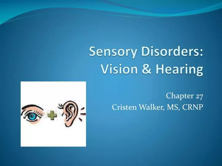
Sensory Disorders: Vision & Hearing. Chapter 27 Cristen Walker, MS, CRNP. Refractive Errors. Refraction = bending of light rays Emmetropia - normal vision Light rays are bent to focus precisely on macula (retina) Ammetropia - refractive error, image not clearly focused on retina

E N D
Sensory Disorders: Vision & Hearing Chapter 27 Cristen Walker, MS, CRNP
Refractive Errors • Refraction = bending of light rays • Emmetropia - normal vision • Light rays are bent to focus precisely on macula (retina) • Ammetropia - refractive error, image not clearly focused on retina • Hyperopia – farsighted, light rays focus behind retina, short eyeball • Myopia – nearsighted, distance vision blurred, long eyeball • Astigmatism - blurred vision w/ distortion, unequal shape of cornea • Presbyopia - older age, loss of ability to focus on close objects, lenses lose elasticity
Refractive errors • Signs & Symptoms • difficulty reading or seeing objects, headaches • Diagnosis: Snellen chart; exam of retina w/ dilation • Treatment: corrective lenses; surgical • Incisional radial keratectomy – incision on cornea to reshape • Photorefractive keratectomy - becoming more common, use of laser to reshape • Intrastromal corneal rings (Intacs) – flexible ring to change cornea shape • for MYOPIA
LASIK • Laser assisted in-situ keratomileusis • Laser used to create thin, circular flap in cornea, folded back, damaged corneal tissue removed, cornea reshaped • Allows light to properly focus on retina • For near-, farsightedness, and astigmatism • Procedure: 5-10 minutes, pt. awake, eye anesthetic used, mild oral sedative, minimal pain
LASIK • Post-operative teaching: • Rest 4-6 hours post procedure • No driving same day • No exercise for 1 week • No eye rubbing • Vision corrected to 20/20 – 20/40 • May still need “reading glasses” (bifocals) • Improved vision within 1-3 days • Complications: Infection, night glare
Disorders of the Eye: Infection • Conjunctivitis - inflammation of conjunctiva
Infection & Inflammation • Blepharitis -inflammation of the eyelid margins (chronic) • Seborrheic • Redness & flaking at base of lashes • Ulcerative • Crusting at lashes, redness of eyes and inflammation of cornea • Tx - long-term daily cleansing • cu-tips & baby shampoo or special eye cleanser • May require antibiotic ointment • Usually caused by staph or effect of rosacea
Infection & Inflammation • Hordeolum - “sty” - external • Small abcess at base of eyelash (red, raised) • Forms in sebaceous gland • Usually resolve spontaneously without treatment • Warm compresses • Chalazion - internal hordeolum • Larger and more problematic • More painful • May require I & D w/ antibiotics
Keratitis • Keratitis - inflammation of the cornea. • Acute or chronic • Caused by infection or irritation • Causes – bacterial conjunctivitis, improper contact lens care, herpes virus, tuberculosis, syphilis, inability of eyelid to close properly • Symptoms: • Pain with eye movement, vision changes, photophobia (sensitivity to light) • Diagnosis: during corneal exam • Treatment: antibiotics or antivirals, artificial tears • If severe, may need corneal transplant (keratoplasty) • Corneal tatoo ?
Keratitis • Treatment • Corneal infections are serious and can result in loss of sight. • May be associated w/ contact lens use • Overnight wearing of contact lens increases risk – can lead to pseudomonas • Must dispose of lenses
Blindness • Complete or almost complete absence of sight. • Often referred to as “visually impaired” to avoid negative connotation • Legal blindness = 20/200 • With corrective lenses
Blindness Causes • Obstruction to ray of light to optic nerve • HTN, diabetes, glaucoma • Disease of optic nerve • Injury/Trauma to occipital lobe (area of sight) • stroke, tumor • Rarely occurs at birth
Blindness • Nursing Interventions • Use normal tone of voice • Alert client when approaching • Orient client to environment • Use a focal point • Allow client to touch objects in room • Promote independence • Provide TV, radio, clocks (oral time) • Assist w/ ambulation – client should walk 1 step behind • Pg. 1258 Textbook: Care of Pt. w/ visual impairment
Cataracts • Opacity (cloudiness) of the lens - may cause loss of vision • Light rays unable to get through • Causes: • Age (>50), sunlight, diabetes, smoking, steroids, alcohol use, infections, congenital defects (rubella), chemical toxins • Painless • Symptoms: • Halos around lights, trouble reading fine print (newspaper), increased sensitivity to glare (sun, car lights), hazy vision, decreased color vision, poor night vision • Looking through a “sheet of falling water”
Cataracts • Surgical removal of cloudy lens, very common procedure • Rare complications – increased ICP, hemorrhage, retinal detachment • Pre-op • Teaching – takes 5-10 minutes, pt. awake (light sedation may be used), eye anesthesia used • Post-op expectations – same day surgery, clear vision within 2 days • Mydriatics may be given pre-op
Cataracts • Post-op care and education • Avoid eye rubbing and straining • Avoid rapid movements; sneezing/vomiting/coughing • Avoid bending, lifting objects >5 lb • Avoid constipation • Use eye drops as prescribed, 2-4 weeks • Use eye shield at bedtime • Contact MD for decrease in vision, severe eye pain, increase in eye discharge
Retinopathy • 2 causes • 1. Diabetes – assoc. w/ excess glucose, constriction of blood vessels & microaneurysms (swell, rupture & hemorrhage) • 2. HTN – retinal vasoconstriction decreases blood supply to retina • Sx: blurred vision, red/black lines or spots, missing areas in field of vision (may be central) • Dx: requires exam of internal eye – annually !
Retinopathy • Tx: Control glucose & BP; prevent further leakage of blood/fluid into retina • Laser surgery • Vitrectomy (vitreous fluid drained & replaced w/ saline) • Cannot reverse vision loss that has occurred, not a cure, may progress • Can progress to retinal detachment and/or blindness
Retinal Detachment • Separation of the retina from the choroid layer allowing fluid to enter the space between the layers. • Caused by: • Hole or tear in the retina • either from degenerative changes or trauma • Fibrous tissue pulling on retina • related to diabetic or hypertensive retinopathy • Accumulation of fluid in the space • from conditions such as HTN, eclampsia, or tumors.
Retinal Detachment • Symptoms: • Gradual or sudden change in vision • loss of peripheral vision or central vision • Depends of portion of retinal involved • flashing lights (colored lights) • floaters or black spots – caused by small hemorrhages • “looking through a veil or cobweb” or “curtain over eyes” • NO PAIN (retina w/o sensory nerves) • Requires prompt referral for treatment (laser reattachment, cryosurgery) • Ages 40-70, increased in Jewish descent (r/t to higher incidence of myopia)
Retinal Detachment • Post op care • Eye patch • Eye shield during sleep to prevent injury • Improvement of vision weeks to months • Flashing lights common for few weeks post op • Antibiotic eyedrops/ointment post op • Pain meds may be prescribed • Wear sunglasses to decrease light sensitivity • No water in affected eye (showers not recommended) • Light activity/rest for several days post op • No sports 3-4 mos. • No driving until 20/40 vision restored
Which finding would the nurse expect while caring for a patient with a detached retina ? • Blurred vision • Pain in the affected eye • Yellow discoloration of the sclera • A sense of a curtain falling across the field of vision
Glaucoma • Abnormal pressure within eyeball • Increased intraocular pressure • Restricts blood flow to retina & optic nerve • Damages optic disc & optic nerve • Damage is usually silent and irreversible • 2nd leading cause of blindness • Sx: loss peripheral vision, reduction in central vision, possible blindness • Can occur at any age • May be genetically predisposed
Glaucoma • Requires lifelong treatment • 3 common types: • AACG – acute angle-closure glaucoma • refers to angle width between cornea & iris • POAG – Primary open-angle glaucoma • chronic • 90% • often inherited • Associated (secondary) glaucoma • w/ DM, HTN, retinal detachment, extreme myopia
Glaucoma • Primary open-angle • Most common • Aqueous fluid behind iris does not flow properly into anterior chamber • No symptoms ?? • Hazy vision, rainbow colored rings around lights, gradual loss peripheral vision, tunnel vision advanced stages • Treatment • Eye drops ** • Laser surgery • Microsurgery – to unblock drainage canals
Glaucoma • Acute angle closure (narrow angle) • Iris pushes against drainage channels of anterior chamber • Drainage canals blocked • Increased intraocular pressure – rises suddenly • Symptoms = severe • Intense eye pain, N/V, sudden vision loss, halos around lights • Medical emergency – prompt treatment • Treatment: laser or surgical iridectomy, to relieve pressure • Failure to treat immediately may cause blindness
Glaucoma • At risk (AACG): • People w/ hyperopia • Asian • Woman >45AACG • AACG: • Requires strict bedrest • Strict avoidance of anticholinergic meds • Mydriatics – dilate pupil
Glaucoma Nursing Interventions: 1. instruct client to wear medic-alert bracelet 2. instruct client to avoid anticholinergic meds 3. Educate re: symptoms *pain in eye, blurred vision, halo w/ lights 4. inform pt. of AACG symptoms – emergency 5. Discuss importance of medication compliance 6. Prevention: low sodium diet, limit caffeine, prevent constipation, decrease stress
A teaching plan for the client with glaucoma should include: • Decrease amount of salt in diet • Decrease fluid intake to control IOP • Avoid reading the newspaper and watching TV • Eye medications will need to be administered for the rest of your life
Macular Degeneration • Leading cause of visual impairment in U.S. • >50. • Deterioration in macula • center of retina • light rays converge, color vision occurs • 2 types • Dry – age related, more common, formation of yellow deposits (drusen), causes drying/thinning • Wet – leaky blood vessels, swelling; more severe • High risk: • >60 yrs, family history of MD, diabetes, smoking, exposure to UV light, caucasian, female, obese, high BP, high cholesterol
Macular Degeneration • Symptoms • blurred vision, distortion straight lines, dark spot in central vision, loss can be progressive (dry) or sudden (wet) • Treatment • Antioxidants/Zinc – may slow progression • Vit. C, Vit. E, Beta Carotene (Vit. A) • Possible laser surgery to seal leaking blood vessels • Not highly effective • Telescopic lens implant