Download
1 / 60
720 likes | 1.15k Vues
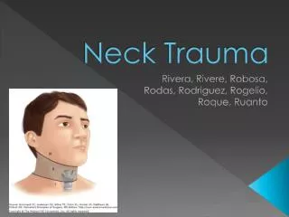
Neck Trauma. Objectives . At the conclusion of this presentation the participant will be able to: Examine the spectrum of neck trauma, the mechanisms of injury and associated injury patterns Define the three zones of the neck used as classifications of injury

E N D
Objectives At the conclusion of this presentation the participant will be able to: • Examine the spectrum of neck trauma, the mechanisms of injury and associated injury patterns • Define the three zones of the neck used as classifications of injury • Identify the appropriate diagnostic modalities used to evaluate patients with neck trauma • Explain the therapeutic interventions in the management of neck trauma • Identify nursing interventions important in caring for patients with neck trauma
Epidemiology • 3500 deaths per year • Mortality rate 2-6% • Blunt mechanism accounts for 5% • Penetrating trauma accounts for most • Zone I injuries are the most lethal
Epidemiology • Commonly injured vessels • Internal jugular vein • Internal carotid artery • Laryngeal and tracheal more common than pharyngeal and esophageal injuries
Blunt Mechanism of Injury • Steering wheel • Assault • Strangulation/Hanging • “Clothes line” injuries • Other (sports, industrial, etc.)
Penetrating Mechanism of Injury • Missile injury (bullet, knife, or other) • Stabbing or lacerations • Impalement • Animal bites
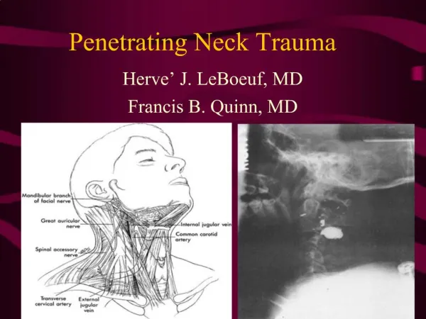
Anatomical Review Fascia Deep cervical fascia Superficial fascia
Structures at Risk Musculoskeletal • Vertebral bodies • Cervical muscles and tendons • Clavicles, 1st and 2nd ribs • Hyoid bone Glandular • Thyroid • Parathyroid • Submandibular • Parotid glands
Structures at Risk Visceral structures • Thoracic duct • Esophagus • Pharynx • Larynx • Trachea
Zones of the Neck • Zone III - Clavicles and sternal notch to cricoid cartilage • Zone II – Cricoid cartilage to the angle of mandible • Zone I – Angle of mandible to base of skull III II I
Zones of the Neck Zone I Zone II Zone III
Zone I • Subclavian vessels • Brachiocephalic veins • Common carotid arteries • Aortic arch • Jugular veins • Esophagus • Lung apices • C- spine/cord • Cranial nerve roots
Zone II • Carotid and vertebral arteries • Jugular veins • Pharynx • Larynx • Trachea • Esophagus • C-spine/cord
Zone III • Salivary and parotid glands • Esophagus • Trachea • Vertebral bodies • Carotid arteries • Jugular veins • Cranial Nerves IX-XII
History and Physical • Gun • Caliper, distance • Knife • Length, angle • Amount of blood loss • Baseline mental status • Baseline motor status • Drug or alcohol use
Key Findings Soft signs Voice change Wide mediastinum Hemoptysis Hematemesis Dysphonia/dysphagia Hard signs • Airway obstruction • Pulsatile bleeding • Expanding hematoma • Unresponsive to resuscitation • Extensive subcutaneous emphysema
Management - Primary Survey • ABCs • Ensure airway is patent • Ensure patient is adequately oxygenating • Control any obvious hemorrhaging • IV access
Airway Considerations Who requires immediate intubation? • Apneic • Comatose • Respiratory compromise • Expanding neck hematoma • Massive subcutaneous emphysema • Massive bleeding in airway
Airway Considerations • “Wait and See” • Avoid excessive bag-valve-mask • Exercise caution with paralytics and sedation • Surgical airway last resort • Cricothyrotomy vs. tracheostomy
Control Bleeding • Local pressure only • No tourniquets • No pressure dressings • No probing or blind clamp placement http://chestofbooks.com
Physical Exam • Violation of the platysma muscle • CNS exam • Obvious hematoma, bleeding
Physical exam • Contusions, lacerations, abrasions to the neck, etc. • Expanding hematomas, obvious bleeding • Hoarseness, stridor, • Subcutaneous emphysema • Hemoptysis, drooling • Dyspnea • Distortion of the normal anatomic landmarks • Mandibular/midface instability
Diagnostic Studies • Chest radiograph • CT and CT angiogram • Laryngeal injury • Tracheal injury • Vessels • Blunt esophageal injury
Diagnostic Studies CT Scan • Can aid in identifying weapon trajectory and structures at risk • Should only be used in stable patients • Gracias et al (2001) found that use of CT scan in stable patients • Saved patients from arteriogram indicated by older protocols 50% of the time • Avoided esophagoscopy in 90% of patients who might otherwise have undergone it
Diagnostic Studies • Laryngoscopy • Bronchoscopy • Esophagoscopy; esophagram • Rigid vs. flexible esophagoscopy • Color flow doppler, duplex ultrasonography • MRA
Diagnostic Studies Arteriogram • Gold standard • Invasive • Complications • Availability varies • Expensive • Contrast load • Simultaneous intervention
Specific Injuries • Vascular • Aerodigestive • Cranial nerves • Thoracic duct
Vascular Injuries in the Neck Physical Exam • External marks • Decreased LOC • Hemiparesis • Hematoma • Hypotension • Dyspnea • Thrill, bruit, pulse not present
Associated Injuries • Le Fort II or III fractures • Basilar skull fracture involving the carotid canal • Diffuse Axonal Injury with GCS < 6 • Cervical vertebral body fracture • Near hanging with anoxic brain injury • Seatbelt abrasion of anterior neck with significant swelling/altered mental status
Primary Diagnostics • CT angiogram of the neck • Chest x-ray indicated in Zone I injuries because of their proximity to the chest • Complete blood count, basic metabolic panel, toxicology and blood alcohol content
Vascular Injury Management • Common carotid: repair preferred over ligation in almost all cases • Internal carotid: Shunting is usually necessary • Vertebral: Angiographic embolization or proximal ligation can be used if the contralateral vertebral artery is intact • Internal Jugular: Repair vs. ligation
Management Summary Vascular Injury • Surgical exploration unstable and stable Zone II • Angiography for Zone I and III • Selective, nonoperative management stable Zone II • Embolization high carotid or vertebral artery • Endovascular stent (pseudoaneurysms) • Anticoagulation blunt carotid/vertebral artery
Aerodigestive Injuries • Airway structures • Trachea • Larynx • Thyroid cartilage • Esophagus • If diagnosis < 24 hours • Poor outcome if diagnosed > 24 hours • Pharyngeal
Tracheal and Laryngeal Injuries Signs of injury • Hoarseness and dysphonia • Hemoptysis • Subcutaneous emphysema in the neck and trunk • Tenderness over the trachea
Primary Diagnostics SubQ air Teeth Laryngotracheal Injury • Plain x-rays • Soft tissue emphysema • Airway compression • Fracture of laryngeal cartilages • CT scan • 3D reconstruction • Endoscopy • Flexible vs. rigid • Bronchoscopy/laryngoscopy Cervical Spine
Management Laryngotracheal Injury • Secure the airway • Early repair • Laryngeal fractures • Thyroid fracture most common • Delay of reduction makes it more difficult and return of normal function unlikely
Esophageal Injury Penetrating • Sharp weapon (knife) • High speed projectile (bullet) • Iatrogenic laceration • Lumen outward injury
Esophageal Injury Blunt • Barotrauma • Blast injuries • Crush injuries • Blow to the neck
Esophageal Injury Signs of Injury • Hematemesis • Odynophagia • Dysphagia • Drooling, hypersalivation • Tracheal deviation • Sucking neck wound • Subcutaneous emphysema • Pain with turning neck
Esophageal Injury Diagnostics Radiographic Findings • Plain films • Air in soft tissue planes • Pneumomediastinum • Leakage of fluid into right pleural space • Contrast swallow • Extravasation is diagnostic • CT scan Laboratory Findings • Markers of inflammatory response • Leukocytosis with left shift • Low oxygen saturations • Acidosis on ABG
Esophageal Injury Diagnostics Helical CT • Expedites diagnosis • Trajectory of missile • Associated injuries
Diagnostics Esophageal Injuries Normal Thoracic Leak