Download
1 / 75
750 likes | 1k Vues
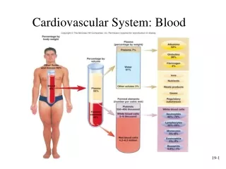
Cardiovascular System Blood and Lymphatic System. Chapter 8 and Chapter 9. Objectives. Discover the functions of the cardiovascular and lymphatic system Identify their location Name the five blood forming organs associated with the circulatory system

E N D
Cardiovascular SystemBlood and Lymphatic System Chapter 8 and Chapter 9
Objectives • Discover the functions of the cardiovascular and lymphatic system • Identify their location • Name the five blood forming organs associated with the circulatory system • Introduce prefixes, root words and suffixes associated with the cardiovascular system
Objectives contd: • Introduce some medical test, lab procedures used in the diagnosis of cardiovascular disease • Introduce some clinical disorders affecting the cardiovascular system
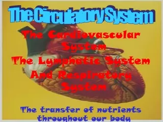
Lets begin!!!!!! • The cardiovascular system is a subset of the circulatory system • It consist of the heart, blood vessels, and blood • The lymphatic system is also a part of the circulatory system • The lymphatic system consist of lymph vessels, and lymph nodes
What is the circulatory system? • The circulatory system carries blood and dissolved substances to and from different places in the body. • The Heart has the job of pumping these things around the body. • The Heart pumps blood and substances around the body in tubes called blood vessels. • The Heart and blood vessels together make up the Circulatory System.
Contd: • Associated with the circulatory system are the blood forming organs: • Spleen • Liver • Bone marrow • Thymus gland • Lymph tissue
Heart: • Main organ of the circulatory system • Weighs less than a pound • Roughly the size of your fist • Lies between your lungs • Found in the thoracic cavity
Heart contd: • Normal beat is about 60 to 100 beats per minute • The apex points down and to the left
Heart contd: • The heart is a pump • Consist of four chambers • Two upper chambers called atria (singular atrium) and two lower chambers called ventricles • The Atrium serves as receiving stations of blood from the body • The ventricles responsible for pumping blood back out into the body
Contd: • There are two sides of the heart. • There are two vertical divisions of the heart. • The top compartments are ATRIUM • The bottom compartments are VENTRICLES • Therefore, there are right and left atrium and right and left ventricles
Contd: • Abbreviations for chambers: Right atrium RA Right ventricle RV Left atrium LA Left ventricle LV
Contd: • The four heart chambers are separated by membranes called septa (plural septum) • The atrium is separated by the Interatrial septum • The ventricles are separated by the Interventricular septum
Heart: • The heart has three distinct layers of tissue. 1. endocardium - deepest layer. 2. myocardium - muscle 3. epicardium - outermost layer
Contd: • The heart is a muscular organ that pumps blood and is enclosed in a membranous sac. This sac allows the heart to beat without friction. • This sac is called the PERICARDIUM. Peri- means “around”, cardium refers to the heart.
Contd: • Valves are the gate keepers of the heart. • They make sure the blood flows in the correct direction • They let a specific amount of blood into each chamber and don’t allow it to flow back wards • There are three valves:
Valves contd: • Bicuspid valve: (mitral) Has two flaps • Situated between the left atrium and left ventricle • Pulmonary and Aortic Semilunar valve: • Pulmonary:Located between the right ventricle and pulmonary artery • Aortic: Located between the left ventricle and the aorta
Valves contd: • Tricuspid valve: Located between the right atrium and the right ventricle • Has three flaps • Keeps blood flowing back and forth • Regulates blood pressure in the heart
Blood vessels: • Series of closed tubes that carry blood from the heart to the tissue and back to the heart. • Made up of arteries, veins, and capillaries • Starts at the heart and spans out through the entire body • These vessels work together to carry blood pumped by the heart through the body
The Blood Vessels • The cardiovascular system has three types of blood vessels: • Arteries (and arterioles) – carry blood away from the heart • Capillaries – where nutrient and gas exchange occur • Veins (and venules) – carry blood toward the heart.
Arterial system: • Arteries: Large blood vessels that carry oxygenated blood away from the heart. • Starts with the Aorta which is the largest artery • Their walls are made up mostly of muscle and elastic tissue • They take the blood to the arterioles then to the capillaries • This is where the exchange of gases (oxygen and carbon dioxide ) take place.
The ARTERY Arteries carry blood away from the heart. the elastic fibres allow the artery to stretch under pressure thick muscle and elastic fibres the thick muscle can contract to push the blood along.
Venous system: • Carry blood back to the atria of the heart • Holds 75% of total blood volume • Begins at the capillary beds, then venules (little veins), then into small, medium and large veins • The veins are the work horse of the vessel system
Veins contd: • They carry oxygen depleted blood back to the heart • Blood is returned to the heart from the upper body through the superior vena cava. • Blood is returned to the heart from the lower body through the inferior vena cava • These veins are the largest veins in the body
The VEIN Veins carry blood towards from the heart. veins have valves which act to stop the blood from going in the wrong direction. thin muscle and elastic fibres body muscles surround the veins so that when they contract to move the body, they also squeeze the veins and push the blood along the vessel.
The CAPILLARY Capillaries link Arteries with Veins they exchange materials between the blood and other body cells. the wall of a capillary is only one cell thick The exchange of materials between the blood and the body can only occur through capillaries.
Circulatory system: • Arteries, arterioles, veins, venules, and capillaries, together with the heart, form the circulatory system. • It can be divided into three types of circulation. • Pulmonary, systemic, and portal
Contd: • Pulmonary System: The right ventricle pumps deoxygenated blood into the pulmonary arteries which then carry the blood to the lungs. Carbon dioxide is released and the uptake of oxygen from the air occurs. Now oxygenated blood returns to the left atrium via the pulmonary veins
Contd: • Systemic system: Oxygenated blood from the lungs return to the heart through the pulmonary veins, flows into the left atrium and then into the left ventricle, which then pumps the blood through the aorta
Contd: • Portal system: The deoxygenated blood from the capillaries of the gastrointestinal tract drains into the portal vein which, instead of going directly back to the heart, leads to the liver. This allows the liver to take up the nutrients that were extracted by the intestines from food. The liver also neutralizes some toxins taken up by the intestines. Blood from the liver drains via the hepatic veins into the inferior vena cava and then the right side of the heart.
Functions of the circulatory system • Transport: • H2 O and nutrients from the intestine to the cells or to a storage site. • O2 from the respiratory organ to the cells and CO2 from the cells back to the respiratory organ. • hormones from endocrine glands. • toxic or waste molecules to the excretory organ.
Contd: • Protection: • from foreign invaders (immune system) • of itself from loss of blood (clotting mechanism) • Body temperature: • Blood vessels dilate to dissipate heat • Or Constricting to retain heat
Contd: • Buffering: • Blood proteins provide an acid base buffer • This maintains optimum pH of the blood
Conduction system • Also known as the cardiac cycle • It is controlled by the hearts natural pace maker the sinoatrial ( SA) node. • The SA node generates electrical impulses and conducts them throughout the muscle of the heart, stimulating the heart to contract and pump blood
Contd: • Atypical muscle fibers at the junction of the superior vena cava and right atrium • Its electrical signals normally cause the atria to contract at a rate of 60 to 100 times per minute. • The electrical current is then passed to the atrioventricular (AV) node
Contd: • The AV Node acts as the primary electrical connection between the atria at the top of the heart and the ventricles in the bottom of the heart • The AV node immediately sends the electrical impulse to the AV bundle called bundle of his. • Then through to specialized muscle fibers called Purkinje fibers.
The Cardiac Cycle and Heart Sounds • The CARDIAC CYCLE is the events that occur in one complete heartbeat. • The cardiac cycle has 2 phases: 1. contraction of the heart: SYSTOLE 2. relaxation of the heart: DIASTOLE
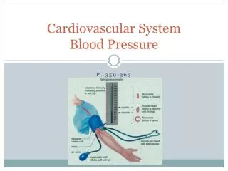
Blood Pressure: • Blood pressure is the force that the blood exerts on the arterial walls • Blood pressure is measured in millimeters of mercury (mm Hg) and is recorded as two numbers usually written one above the other; for example, 120/80. The top number is the systolic number, and the bottom number is the diastolic number. Normal Blood pressure is around 120/80.
Contd: • Systolic blood pressure measures the maximum (highest) pressure in the arteries during the cardiac cycle, which occurs when the heart contracts, or beats, to pump blood. • The systolic blood pressure, marks the beginning of the cardiac cycle, when the heart contracts. • The top number in blood pressure reading
Contd: • Diastolic blood pressure measures the pressure exerted by the heart against artery walls when the heart is at rest. • The diastolic pressure marks the end of the cardiac cycle, when the heart fills with blood and are dilated. • The bottom number in a blood pressure reading
The Cardiovascular System • Pulse is the expansion and contraction of an artery. • Blood pressure is a measurement of the amount of pressure exerted on the walls of the vessels.
FYI: • High blood pressure, also called hypertension, is defined as 140/90 mm Hg or higher. It is when there is high pressure (tension) in the arteries. • Low blood pressure, or hypotension, occurs when blood pressure during and after each heartbeat is much lower than usual. This means the heart, brain, and other parts of the body do not get enough blood
Arteriosclerosis: Atherosclerosis • Arteriosclerosis is plaque or cholesterol, platelets, fibrin and other substances on the arterial walls (artery. • Atherosclerosis is a condition in which fatty material collects along the walls of arteries. This fatty material thickens, hardens (forms calcium deposits), and may eventually block the arteries. • Atherosclerosis is a type of arteriosclerosis. Thetwo terms are often used to mean the same thing
Coarctation: • Stricture or narrowing of a vessel • A coarctation can occur anywhere in the aorta, but it is most often found just beyond the point where the aorta sends a branch off to supply the left arm.
Congestive Hear Failure: (CHF) • Congestive heart failure (CHF) is a condition in which the heart's function as a pump to deliver oxygen rich blood to the body is inadequate to meet the body's needs. CHF can be caused by: • diseases that weaken the heart muscle • diseases that cause stiffening of the heart muscles • diseases that increase oxygen demand by the body tissue beyond the capability of the heart to deliver.
Coronary Thrombosis: • Thrombosis is the formation of a blood clot (thrombus) inside a blood vessel, obstructing the flow of blood through the circulatory system. • Coronary Thrombosis is the thrombosis of a coronary artery. This can lead to an Myocardial infarction (MI) (Heart attack)
Transient Ischemic attack (TIA) • TIAs occur when a blood clot temporarily clogs an artery, and part of the brain doesn't get the blood it needs. • A TIA is a "warning stroke" or "mini-stroke" that produces stroke-like symptoms but no lasting damage.
Abbreviations: • ALL acute lymphocytic leukemia • a fast-growing cancer in which the body produces a large number of immature white blood cells (lymphocytes). • AMI acute myocardial infarction • ASHD arteriosclerotic heart disease • MI Myocardial infarction
Contd: • CVA cerebrovascular accident • The sudden death of some brain cells due to lack of oxygen when the blood flow to the brain is impaired by blockage or rupture of an artery to the brain. A CVA is also referred to as a stroke. • O2 oxygen • RBC Red blood cell
Contd: • ASDarterial ( atrial) septal defect • An atrial septal defect is an opening in the atrial septum, or dividing wall between the two upper chambers of the heart known as the right and left atria. ASD is a congenital (present at birth) heart defect. • BP Blood pressure • CBC Complete blood count
Contd: • CABG Coronary artery bypass graph • During a coronary artery bypass graft, blood flow is rerouted through a new artery or vein that is grafted around diseased sections of a coronary artery to increase blood flow to the heart muscle tissue.