Download
1 / 15
150 likes | 316 Vues
High-Resolution Determination of Coral Reefs using Fluorescence Imagery Laser Line Scanner (FILLS). Laser Line Scanning. Electro-optic imaging technique Application of lasers for mapping of benthic environment Lasers allow for higher image resolution Superior object/organism recognition

E N D
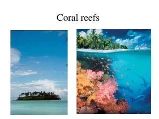
High-Resolution Determination of Coral Reefs using Fluorescence Imagery Laser Line Scanner (FILLS)
Laser Line Scanning • Electro-optic imaging technique • Application of lasers for mapping of benthic environment • Lasers allow for higher image resolution • Superior object/organism recognition • More sophisticated than traditional technologies • Trawl • Direct Observation
FILLS • Seeks to: • map coral reef bathymetry • Evaluate degree to which organisms are present • Generating high-resolution image of an entire reef
FILLS • A towed system (behind a vessel or attached to a submersible) • Emits a swath blue-green (488 nm) light • Inverse relationship between width of the swath and resolution of image • Determined by altitude of instrument • Limited by water clarity
FILLS distance limitations • Laser Line Scanning (LLS) results have been of sufficient quality within five to six beam attenuation lengths • Distance over which beam intensity is reduced by a factor of 1/e, or ~63%, by absorption and scattering
FILLS • Four sensors are equipped with a 488 nm filter to exclude reflected light • Each sensor channel generates a distinct black & white image of the area • Channels measure: • 488 nm (reflectance) • 520 nm (green) • 580 nm (orange) • 685 nm (red)
Raw FILLS Results 488 nm 520 nm 580 nm 685 nm reflectance green orange red composite
FILLS • Each channel is mapped to display its respective color, and the four images are combined to display a composite • Significant because different organisms fluoresce at different wavelengths, meaning different groups and species may have fluorescent signatures (therefore, the use of distinct sensors)
FILLS • Coral reef organisms were able to be distinguished significantly based on fluorescence alone due to symbiotic zooxanthellae, exhibiting a maximum fluorescence at ~685 nm (red) due to Chlorophyll a • Macroalgaes, Sediments, Sponges, and non-fluorescent organisms can likewise be distinguished for different reasons
FILLS • Data is analyzed using an algorithm which defines relationships between adjacent pixels the resulting objects, in order to determine to which taxonomic group an organism belongs
FILLS data check • Composite images are then printed on waterproof paper, and divers are able to conduct direct observations to verify the algorithm calculations • With few corrections, the following image results:
Where: • White = corals, anemones, zooanthids • Red/brown = gorgonians • Green = sand/substrate • Blue = red algal turf • Black = shadows/fish & Calyspongia vaginalis • Purple = Xestospongia muta • Pink = Unknown Near left: image detail, with barrel sponge Xestospongia muta at bottom left, with algae growing in the center of the sponge (indicated by the red) Far Left: mottled orange are zooanthid colonies (Palythoa caribaeorum)
FILLS Shortcomings • Small-scale data inconsistencies • Divers are either more or less accurate • Cost of operations • Until direct observation becomes unnecessary, this method is cost prohibitive • Common presence of Chlorophyll a • This pigment fluoresces most brightly at ~685 nm (red), and makes it difficult to distinguish organisms with similar morphology and fluorescent signal
FILLS Utility • Coral reef diversity and organism frequency • Population dynamics over time • Track & observe coral bleaching on colonies and entire reefs
Other LLS Applications • Identify aggregations of bottomfish • Potential bottomfish nursery areas • Various coral communities • Airplane wreckage location and imaging • e.g.TSA flight 800 • Still more affordable and accurate than traditional still or video photography • Can map greater area in less time, with less disturbance to inhabitants of the ecosystem