Download
1 / 15
150 likes | 248 Vues
Figure 10.1A. DNA is the carrier of genetic information. Head. DNA. Tail. Tail fiber. Figure 10.1B. Empty protein shell. Radioactive protein. Phage. The radioactivity is in the liquid. Bacterium. Phage DNA. DNA. Batch 1: Radioactive protein labeled in yellow. Centrifuge. Pellet. 3.

E N D
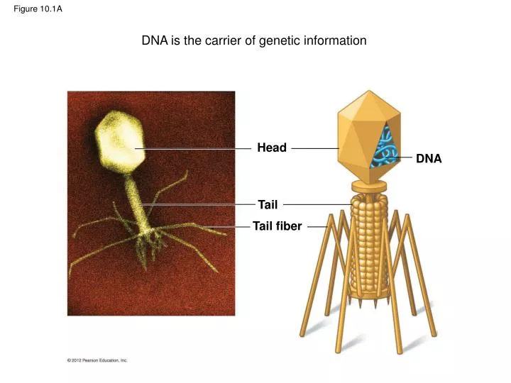
Figure 10.1A DNA is the carrier of genetic information Head DNA Tail Tail fiber
Figure 10.1B Emptyprotein shell Radioactiveprotein Phage The radioactivityis in the liquid. Bacterium PhageDNA DNA Batch 1:Radioactiveproteinlabeled inyellow Centrifuge Pellet 3 1 2 4 Batch 2:RadioactiveDNA labeledin green RadioactiveDNA Centrifuge The radioactivityis in the pellet. Pellet
Figure 10.2A T A C G Sugar-phosphatebackbone T A Phosphategroup C G T A C G A Nitrogenousbase A G Nitrogenous base(can be A, G, C, or T) Covalentbondjoiningnucleotides A T Sugar G C T A C C C C T A C G T A DNAnucleotide Thymine (T) T T A DNAdouble helix T Phosphategroup G G Sugar(deoxyribose) DNA nucleotide G G Two representationsof a DNA polynucleotide
Figure 10.2B Cytosine (C) Guanine (G) Thymine (T) Adenine (A) Pyrimidines Purines
Figure 10.3D Hydrogen bond Base pair Computermodel Ribbonmodel Partial chemicalstructure
Central Dogma: information flow from genes to proteins The term ‘central dogma’ is a hypothesis attributed to Francis Crick, the co-discoverer of the DNA double helix structure DNA Interactive (www.dnai.org)
Figure 10.4A_s1 A T C G G C A T T A A parentalmoleculeof DNA
Figure 10.4A_s2 T A T A A T C G C G G C G C G C C A A T T A Freenucleotides T A T A A parentalmoleculeof DNA The parental strandsseparate and serveas templates
Figure 10.4A_s3 T A T A T A T A A T C G C G C G C G G C G C G C G G C C C A A T A T T A A T Freenucleotides T A T A T A T A A parentalmoleculeof DNA The parental strandsseparate and serveas templates Two identicaldaughter moleculesof DNA are formed
Figure 10.4B AT G C A T Parental DNAmolecule A T T A C G G C T Daughterstrand A C G C G Parentalstrand C G T C G A A T C G A T T A C G G C A T T A T A G C AT Daughter DNAmolecules
Figure 10.5A ParentalDNAmolecule Parental strand Origin of replication Daughter strand “Bubble” TwodaughterDNAmolecules
Figure 10.9 TerminatorDNA RNA polymerase DNA of gene PromoterDNA Initiation 1 Area shownin Figure 10.9A Elongation 2 GrowingRNA Termination 3 CompletedRNA RNApolymerase
Figure 10.10 Intron Exon Exon Intron Exon DNA TranscriptionAddition of cap and tail Cap RNAtranscriptwith capand tail Introns removed Tail Exons spliced together mRNA Coding sequence NUCLEUS CYTOPLASM
Figure 10.13A Start of genetic message Cap End Tail