Download
1 / 5
50 likes | 123 Vues
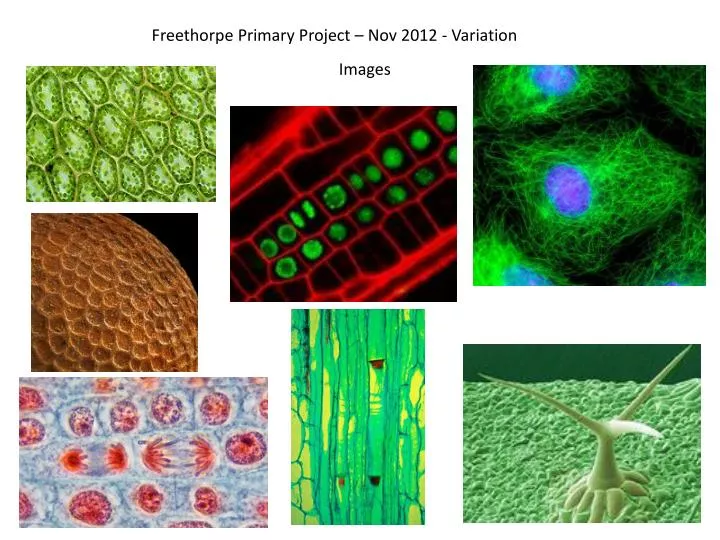
Freethorpe Primary Project – Nov 2012 - Variation. Images. All Science photo library so no permission to use as yet!. Light micrograph showing a section through a leaf. Each cell contains several round, green vescicles which are known as chloroplasts Credit: JOHN DURHAM/SCIENCE PHOTO LIBRARY.

E N D
All Science photo library so no permission to use as yet! Light micrograph showing a section through a leaf. Each cell contains several round, green vescicles which are known as chloroplasts Credit: JOHN DURHAM/SCIENCE PHOTO LIBRARY Black mustard seed Credit: POWER AND SYRED/SCIENCE PHOTO LIBRARY Mitosis in plant root. Confocal light micrograph showing epidermal cells of a plant root. The cell walls can be seen in red. Chromatin in the nucleus (green) of one of the cells has separated (centre left) during the late anaphase stage of mitosis (nuclear division). Fluorescent dyes have been used to highlight the cell structures and proteins. This root is from the Arabidopsis thaliana plant. Credit: DR JOHN RUNIONS/SCIENCE PHOTO LIBRARY Cell division. Immunofluorescent light micrograph of a human epithelial cell (centre) during the interphase stage of mitosis. Mitosis is the cycle of replication and division by which new body cells are formed. Here, the chromosomes of the parent cell (blue) have begun to duplicate and pair up, and the microtubules (green) have formed. Credit: DR TORSTEN WITTMANN/SCIENCE PHOTO LIBRARY Onion (Allium) root tip mitosis Credit:WIM VAN EGMOND/VISUALS UNLIMITED, INC. /SCIENCE PHOTO LIBRARY Plant phloem tissue. Light micrograph of a longitudinal section through tissue from a sunflower (Helianthus) plant, showing sieve tubes (horizontal structures) and sieve plates (red areas) in phloem tissue Credit: BIOPHOTO ASSOCIATES/SCIENCE PHOTO LIBRARY Trichome on the leaf of (Arabidopsis thaliana). Credit: DR. STANLEY FLEGLER/VISUALS UNLIMITED, INC. /SCIENCE PHOTO LIBRARY
Science SAW 051.jpg SAW 037.jpg SAW 040.jpg SAW 015.jpg SAW 038.jpg Mashing up bananas for DNA extraction SAW 044.jpg
Writing Writer & storyteller, Kelly Kanayama SAW 029.jpg
Art Art work 4 for SAW project .jpg Art work for SAW project 2.jpg Art work for SAW project 3.jpg Art work for SAW project .jpg