Download
1 / 48
500 likes | 790 Vues
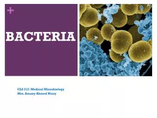
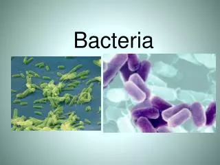
Bacteria. History of Microbiology. 1664: Robert Hooke - microscope 1684: Antoni van Leeuwenhoek - microorganisms 1798: Edward Jenner - smallpox vaccination 1864: Louis Pasteur - spontaneous generation 1884: Robert Koch - Koch’s postulates 1889: Martinus Beijerink - concept of virus

E N D
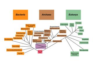
History of Microbiology • 1664: Robert Hooke - microscope • 1684: Antoni van Leeuwenhoek - microorganisms • 1798: Edward Jenner - smallpox vaccination • 1864: Louis Pasteur - spontaneous generation • 1884: Robert Koch - Koch’s postulates • 1889: Martinus Beijerink - concept of virus • 1929: Alexander Fleming - discovery of penicillin • 1977: Carl Woese - discovery of Archaea • 1981: First reports of AIDS • 1983: Luc Montagnier - discovery of HIV • 1995: Craig Venter - complete genome sequence
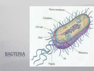
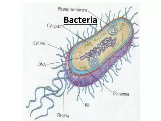
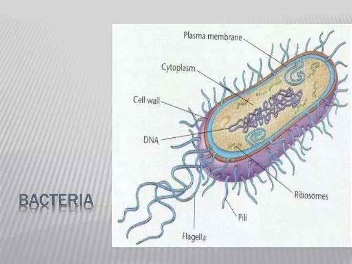
Characteristics • Prokaryotes • Microscopic • (Eukaryotic cells are at least 10x bigger) • Unicellular • DNA is a single circular piece of DNA • Asexual Reproduction • Binary Fission • Metabolism • Aerobic • Anaerobic
Genetic Exchange • Conjugation –transfer DNA through contact • Transformation – acquire DNA from dead bacteria • Transduction – DNA is transferred from one bacteria to another using a virus (genetic engineering)
http://highered.mcgraw-hill.com/sites/0072556781/student_view0/chapter13/animation_quiz_2.htmlhttp://highered.mcgraw-hill.com/sites/0072556781/student_view0/chapter13/animation_quiz_2.html http://highered.mcgraw-hill.com/sites/0072556781/student_view0/chapter13/animation_quiz_3.html
allow them to withstand drought, high temps., lack of food, etc. endospores
Gram Staining • Gram + simple walls, large amount of peptidoglycan • Gram - less peptidoglycan, outer membrane contains lipopolysaccharides which are often toxic and provides additional protection more resistant to antibiotics • Many antibiotics (penicillens) inhibit synthesis of cross links in peptidoglycan and prevent formation of a functional wall Gram positive Gram negative http://highered.mcgraw-hill.com/sites/007337525x/student_view0/exercise9/gram_stain.html
Gram Positive Organisms • Aerobic, Gram-positive cocci • Staphylococcus aureus (fig 1, 2, 3, 4) • Staphylococcus epidermidis (fig 1) • Staphylococcus sp. (Coagulase-negative)(fig 1) • Streptococcus pneumoniae (Viridans group)(fig 1, 2, 3) • Streptococcus agalactiae (group B)(fig 1) • Streptococcus pyogenes (group A)(fig 1, 2) • Enterococcus sp.(fig 1, 2, 3) • Aerobic, Gram-positive rods • Bacillus anthracis (fig 1, 2) • Bacillus cereus (fig 1, 2) • Bifidobacterium bifidum (fig 1) • Lactobacillus sp. (fig 1, 2) • Listeria monocytogenes (fig 1, 2) • Nocardia sp.(fig 1, 2) • Rhodococcus equi (coccobacillus)(fig 1) • Erysipelothrix rhusiopathiae (fig 1) • Corynebacterium diptheriae (fig 1, 2) • Propionibacterium acnes (fig 1) • Anaerobic, Gram-positive rods • Actinomyces sp. (fig 1, 2) • Clostridium botulinum (fig 1) • Clostridium difficile (fig 1) • Clostridium perfringens (fig 1, 2, 3) • Clostridium tetani (fig 1, 2) • Anaerobic, Gram-positive cocci • Peptostreptococcus sp. (fig 1)
Gram Negative Organisms • Aerobic, Gram-negative cocci • Neisseria gonorrhoeae (fig 1, 2, 3, 4) • Neisseria meningitidis (fig 1; false color of the bacterium., 2) • Moraxella catarrhalis (fig 1) • Anaerobic, Gram-negative cocci • Veillonella sp. (fig 1) • Aerobic, Gram-negative rods • Fastidious, Gram-negative rods • Actinobacillus actinomycetemcomitans (fig 1) • Acinetobacter baumannii(fig 1 really A. calcoaceticus) • Bordetella pertussis (fig 1, 2) • Brucella sp. (fig 1) • Campylobacter sp.(fig 1) • Capnocytophaga sp.(fig 1,2) • Cardiobacterium hominis (fig 1) • Eikenella corrodens (fig 1) • Francisella tularensis (fig 1,) • Haemophilus ducreyi (fig1,2) • Haemophilus influenzae (fig 1, 2) • Helicobacter pylori (fig 1, 2, 3, 4) • Kingella kingae (fig ) • Legionella pneumophila (fig 1, 2, 3) • Pasteurella multocida (fig 1) • Enterobacteriaceae (glucose-fermenting Gram-negative rods) • Citrobacter sp. (fig 1) • Enterobacter sp. (fig 1) • Escherichia coli (fig 1, 2) • Klebsiella pneumoniae (fig1, 2) • Proteus sp. (fig 1) • Salmonella enteriditis (fig 1) • Salmonella typhi (fig 1) • Serratia marcescens (fig 1, 2) • Shigella sp. (fig 1) • Yersinia enterocolitica (fig 1) • Yersinia pestis (fig 1, 2) • Oxidase-positive, glucose-fermenting Gram-negative rods • Aeromonas sp. (fig 1) • Plesiomonas shigelloides (fig 1) • Vibrio cholerae (fig 1, 2) • Vibrio parahaemolyticus (fig 1) • Vibrio vulnificus (fig 1) • Glucose-nonfermenting, Gram-negative rods • Acinetobacter sp. (fig 1) • Flavobacterium sp. (fig 1) • Pseudomonas aeruginosa (fig 1, 2) • Burkholderia cepacia (fig 1) • Burkholderia pseudomallei (fig 1) • Xanthomonas maltophilia or Stenotrophomonas maltophila(fig 1) • Anaerobic, Gram-negative rods • Bacteroides fragilis (fig 1) • Bacteroides sp. (fig 1) • Prevotella sp. (fig 1) • Fusobacterium sp. (fig 1,2) • Gram-negative spiral • Spirillum minus (minor)- (fig 1)
Nutrition • Autotrophic • Photosynthetic • Chemoautotrophic (nitrogen fixers) • Heterotrophic • Decomposer • Parasitic (Treponema pallidum)
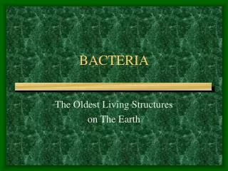
Survival of the Fittest!!! Bacteria have been around for 3.5 billion years!! How???? • Cell Walls • Capsules (surrounds cell wall) • Asexual Reproduction, but can still acquire other genes • Inhabit every place on Earth
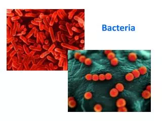
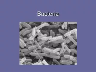
Shapes • Coccus : Spheres • Bacillus : Rods • Spirillum : Spirals • Arrangements • Strept : Chains • Staph : Clusters • Diplo : Pairs Bacteria are Classified according to Shape and arrangement of cells
Important Recyclers in environment • Nitrogen cycle
Bacteria can produce chemicals • Acetone, Butanol
Bacteria are used to make food • Pickles, buttermilk, cheese, sauerkraut, olives, vinegar, sourdough bread, beer, wine
Bacteria cause disease 1. Produce toxins: example (Clostridium botulinum) • Endotoxins : • part of cell wall of gram – bacteria (lipids) • Dead bacteria release toxins • Exotoxins • Gram + • Very toxic • Easily transported throughout body 2. Metabolize their host (Mycobacterium tuberculosis) Bacterial Diseaes
Streptococcus Bacteria • Natural reservoir: humans • Scarlet fever • Pharyngitis • Pneumonia • Cause more illness than any other bacteria group • Gram + chains
ViBriocholerae • Natural reservoir: humans • Cholera • Caused by poor sanitation • Common in Asia and Africa • Gram -
Salmonella Bacteria • Gut of mammals • Cause of gastrointestinal diseases • Typhoid Fever • Gram -
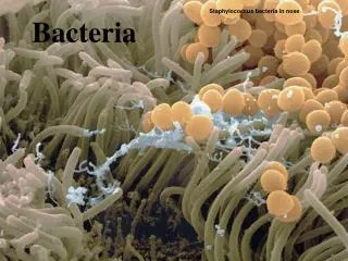
Staphylococcus Aureus • Natural reservoir: humans • Normally inhabits nose, skin and growing in cured meats (ham) • Most common cause of food poisoning • Produces toxins • Problematic in hospitals • Antibiotic resistance • Gram + spheres
Enterobacter Cloacae • Natural reservoir: mammals • Urinary tract infections and respiratory infections • Normal gut flora • Common in water sewage and soil • Used to control plant diseases • Gram - rods
Haemophilusinfluenzae • DOES NOT cause flu • Inhabits mucous membranes of Upper respiratory tract and mouth • Causes meningitis ear aches, bronchitis and pneumonia (mainly affects 5 yrs and under) • Opportunistic pathogen • Gram -
Yersinia pestis • Black Death or Plague of medieval Europe • Claimed 1/3 of European population in 1300’s • Fleas from rats and squirrels transmit • Gram – rod • CDC id as biological warfare agent https://www.youtube.com/watch?v=kScxc9DPrnY
Clostridium Botulinum • Soil • Causes Botulism • Secretes neurotoxins which can destroy, paralyze or damage nerve cells • Initial symptoms: nausea, vomiting, diarrhea • Death results from respiratory distress
How can Botulism be Contracted? For example, if a low-acid food, such as green beans, is canned improperly (not canned under pressure or improperly canned using a pressure canner), C. botulinumbacteria and other bacteria present will be destroyed by the boiling of water and food, but the C. botulinum spores will not be destroyed. The canning process will remove the oxygen from the jar, creating a low-oxygen environment that is will allow the spores to grow into active bacteria. When the jars are stored at room temperature, the spores can germinate and produce the toxin. However, the toxin is sensitive to heat and can be destroyed if the food in question is boiled for 10 minutes (longer at high altitudes).
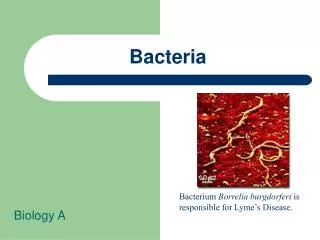
Treponemapallidum • Helical bacterium moves in corkscrew manner • Causes syphilis: STD http://www.microbiologybytes.com/video/Tpallidum.html
Neisseria gonorrhaeae • Gram – spherical pairs • Causes gonorrhea • Fimbriae enable the organism to attach to mucous membranes of vagina and urethra of penis