Download
1 / 46
490 likes | 727 Vues
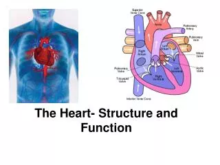
Structure of the Heart. 2009-07-21. Facts, location, & orientation. Oblique orientation Apex points inferiosinister (down and left) 5 th intercostal space Base is superior near origins of great vessels 2 nd intercostal space 2/3 lies left of the midline For the most part

E N D
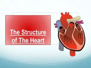
Structure of the Heart 2009-07-21
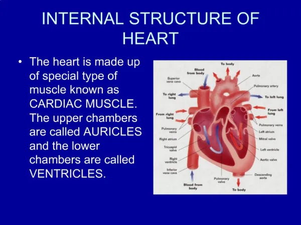
Facts, location, & orientation • Oblique orientation • Apex points inferiosinister (down and left) • 5th intercostal space • Base is superior near origins of great vessels • 2nd intercostal space • 2/3 lies left of the midline • For the most part • Anterior/inferior aspect of the heart • right atrium/ventricle • Posterior/superior aspect • left atrium/ventricle
Mediastinum Mediastinum Superior Inferior Anterior Middle Posterior Heart
Structure of the Heart – Coverings • Fibrous pericardium • Serous pericardium • Parietal pericardium • Visceral pericardium
Sulci of the heart • Coronary sulcus • Atrioventricular sulcus • Circumvents the heart • Interventricular sulcus • Anterior • Posterior
Great vessels • Aorta • From left ventricle • Pulmonary trunk • Originates anterior to the aorta from right ventricle • Superior Vena Cava • Inferior Vena Cava • Both empty into right atrium
Transverse pericardial sinus Oblique pericardial sinus
Aorta • Ascending aorta • Right and left aortic sinuses • Arch of aorta • Begins/ends at T4/T5 or sternal angle level • Brachiocephalic a. • Left common carotid a. • Left subclavian a. • Thoracic aorta • Lies anterior to trachea
Ligamentum arteriosum • Remnant of embryonic ductus arteriosus • Attaches aortic arch superiorly to pulmonary trunk/left pulmonary artery inferiorly • Identification point for Left recurrent laryngeal nerve
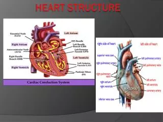
Chambers Right atrium Right ventricle Left atrium Left ventricle Valves Leaflet valves Tricuspid Bicuspid (mitral) Cusped (semilunar) valves Aortic Pulmonic Heart Features
Right atrium • Auricle (ear) • Pectinate muscles (rough) • Sinus venarum (smooth) • Crista terminalis • Division between rough to smooth • Openings (ostia) • SVC/IVC/Coronary sinus • Fossa ovalis • Foramen ovale in fetus • Limbus
Right atrium “valves” • Superior vena cava • No valve • Inferior vena cava • Eustachian valve • Incompetent in adult, directs IVC blood though Foramen ovale in fetus • Coronary Sinus • Thebesian valve • Prevents backflow into coronary sinus during atrial systole
Left atrium • Ostia of 4 pulmonary veins • 2 superior • 2 inferior • Auricle
Right ventricle • Most anterior aspect of heart • Tricuspid valve (RA-RV) • Anterior/Posterior/Septal cusps (leafs) • Papillary muscles • Connected to cusps via Chordae tendinae • Contract to prevent Tricuspid valve regurgitation • Named same as cusps • Trabeculae carnae • Moderator band
Left ventricle • Trabeculae carnae • Bicuspid (mitral) valve • Anterior/Posterior cusps • Papillary muscles • Chordae tendinae • Usually a greater number than the right, due to the increased pressures and strength necessary to prevent regurgutation
Pulmonic valve • From RV to pulmonary trunk • Lies just anterior to aortic valve • 3 semilunar cusps • Anterior • Right • Left
A P
Aortic valve • Posterior to pulmonic valve • Just superior lies the Sinus of Valsalva • Helps to dampen aortic outflow and prevent cusps from adhering to walls of aorta • 3 cusps • Posterior (non-coronary) cusp • Right • Left • Just superior to right and left cusps in the Sinus of Valsalva are the openings of the right and left coronary arteries, respectively
Heart Valves • Tricuspid valve • RA – RV • Bicuspid valve • LA – LV • aka “Mitral valve” • Aortic valve • LV – aorta • Pulmonic valve • RV – pulmonary trunk
Right coronary blood supply • Right coronary artery • Originates from ostia in right aortic sinus • Superior to right aortic cusp • Travels in right coronary (AV) sulcus • Branches • Right marginal arteries (acute marginal aa) • Posterior interventricular a. (in post. IV sulcus) • Sinoatrial nodal a. • Atrioventricular nodal a.
Left coronary blood supply • Left coronary artery • Originates from ostia in left aortic sinus • Superior to left aortic cusp • Branches • Left anterior descending (LAD) or anterior interventricular a. (lies in anterior IV sulcus) • Septal branches. • Diagonal branches • Left marginal aa. (Obtuse marginal aa.) • Left circumflex a.
Dominance • Defined by branch that gives rise to posterior interventricular a. • Right (80%) • From right coronary a. • Left (15%) • From left circumflex a. • Co-dominance (5%)
Venous drainage of the heart • Coronary sinus • Lies in coronary (AV) sulcus on posterior • Opens directly to right atrium • All venous drainage of the heart eventually flows here • Great cardiac vein • With LAD in anterior IV sulcus • Left marginal vein • Middle cardiac vein • With posterior interventricular a. • Small cardiac vein • With right coronary a. • Right marginal vein • Oblique vein (LA) • Posterior vein of the left ventricle
Heart Valves – Transverse Section Tricuspid Aortic Semilunar Bicuspid Left Ventricle