Download
1 / 21
230 likes | 1.07k Vues
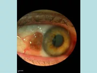
Pterygium Surgery with Sutured Conjunctival Autografts. David S. Rootman, MD, FRCSC Associate Professor, University of Toronto. Why Sutures When We Can Glue?. More secure fixation of autograft Less likely to dislocate Stretches out a smaller autograft Long track record

E N D
Pterygium Surgery with Sutured Conjunctival Autografts David S. Rootman, MD, FRCSC Associate Professor, University of Toronto
Why Sutures When We Can Glue? • More secure fixation of autograft • Less likely to dislocate • Stretches out a smaller autograft • Long track record • No bovine or foreign blood products • Less messy • Good suturing practice • Less expensive
Disadvantages of Sutures • Takes longer • More difficult • Sutures irritate patient • More likelihood of bleeding • May have to remove sutures
Seven Principles of Procedure Smooth partial keratectomy Release of Tenon’s capsule constriction Thin autograft Alignment of graft Secure suturing of graft Bandage contact lens
Keratectomy • As described by Richard Abbott • 7-0 Silk fixation suture • Inject under pterygium with 1% xylo with epi • Angled cut at anterior edge of pterygium • Smooth dissection in anterior stroma • Removal of all scar tissue on cornea • Similar to making a phaco scleral tunnel
Release of Tenon’s Capsule • Do not over dissect • Release medial rectus on both sides • Allow conjuctiva to slide back to caruncle • Mark extent of excision • Minimal removal of conjunctiva • Stay away from caruncle
Autograft harvesting • Measure area of resection • Pull eye downward • Use Gentian violet to mark area • Central mark to help orientation • Rhomboidal shape, wider at posterior edge • Make graft as thin as possible
Alignment of Graft Slide conjunctiva on cornea, Tenon up Appose limbal cells to limbus at site of excision Secure with 10-0 monofilament vicryl Turn graft over after secured at limbus
Suturing of Autograft • Secure in all four corners with scleral bite • Avoid medial rectus to minimize bleeding • Close nasal conjuntiva to conjunctiva of graft, no scleral bite here • Close edge to edge superiorly and inferiorly • No exposed Tenon capsule, prevents granuloma • Do not advise running suture
Bandage Contact lens • Apply at end of procedure • More comfort for patient • No patch needed • Leave on for 2 weeks • Lessens chance of Dellen • Steroids for 6 weeks qid or until eye white
Complications • Recurrence • Inflammation • Melting (more common with Mito C) • Infection • Bleeding • Dellen • Granuloma
Conclusions • Pterygium excision with conjunctival flap is a good procedure with low complication rates • Using sutures is a good alternative compared with tissue glue