Download
1 / 23
250 likes | 705 Vues
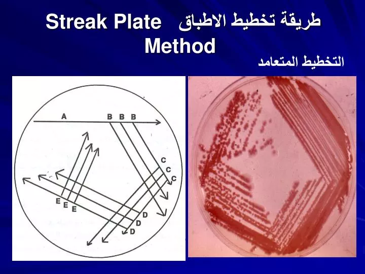
طريقة تخطيط الاطباق Streak Plate Method. التخطيط المتعامد. Streak Plate Method. Gram Stain. spread as a thin layer. One drop of water + Loop of Bacteria. 1-allowed to air dry. smear. 2-fixed. clean glass slide. 2. Saturate the smear with crystal violet for 1 minute.

E N D
طريقة تخطيط الاطباقStreak Plate Method التخطيط المتعامد
Gram Stain spread as a thin layer One drop of water + Loop of Bacteria 1-allowed to air dry smear 2-fixed clean glass slide 2. Saturate the smear with crystal violet for 1 minute. 3. Rinse the slide gently with water. 4. Saturate the smear withiodine for 30 Second. 5. Rinse the slide gently with water. 6. Decolorize (alcohol) 7. Rinse the slide gently with water. 8. Saturate the smear with safranin for 1 minute. 9. Rinse the slide gently with water. 10. Carefully blot the slide dry with bibulous paper. 11. Observe the slide under the microscope
Gram staining Results Gram positive bacteria will stain purple. Gram negative bacteria will stain red/pink. Gram positive bacteria have a thick cell wall made of peptidoglycan, whereas gram negative bacteria have thin layer of peptidoglycan.
Cells Description under the microscope 1- Cell response : GV + or GV-
Cells Description under the microscope 2- Cell shape and arrangement : Cocci: single- Diplo – Grape like-Strepto
Cells Description under the microscope Road: Short or long Single or Chain
Catalase test H2O2 Catalase H2O + O2 (gas, ↑) Staphylococci • The catalase test is distinguished streptococci from staphylococci • flood culture with drops of 3% H2O2 • Catalase-positive cultures bubble at once • The test should not be done on blood agar because blood itself will produce bubbles
Species of Satphylococci • Three species of staphyloccoci have medical importance: • S. aureus: Pathogenic & commensally found in nose (nares) • S. epidermidis:non pathogenic & common commensals in nares & skin • S. saprophyticus: Cause UTI in female & occasionally commensally found skin
Coagulase Test Principle: • This test used to differentiate between S. aureus (CPS) & other Staphylococcus species (CNS) Fibrinogen (Plasma) Coagulase Fibrin (Clot)
General characters: Gram Positive Cocci Grape-like Non Motile Non Spore Forming Non Capsulated Non Fastidious Facultative Anaerobes Fermentative Catalase positive Staphylococci
Laboratory diagnosis of Staphylococcus • Gram Stain: • Gram Positive Cocci, arranged in cluster • Culture: 1-Blood agar , 2- MSA media • Blood agar (Non-Selective Media) • Coagulase Positive Staphylococci are Pigmented & hemolytic • Coagulase Negative Staphylococci are non-pigmented & non-hemolytic
MSA is selective differential medium for staphylococci • It contains: NaCl (7.5%), Mannitol, & Phenol Red • The cause of selectivity due to presence of high salt concentration • The cause of differential because contains mannitol (sugar) and phenol red (pH indicators turns yellow in acidic pH and turns red in alkaline pH).
Coagulase Test • The tube coagulase test (Free): • Procedure: • Mix 0.1 ml of culture + 0.5 ml of plasma • Incubate at 37C for 4 h • Observing the tube for clot formation • Any degree of clotting constitutes a positive test • Advantage • More accurate • Disadvantage • Time consumed S. epidermidis S. aureus
Coagulase Test • Two Methods: • The slide Method • Tube Method • The slide coagulase test • Used to detect bound coagulase or clumping factor • Add one drop heavy bacterial suspension and one drop of plasma on clean slide • Mixing well and observing for clumping within 10 seconds • Advantage • Rapid diagnosis • Disadvantage • Less accurate
Deoxyribonuclease (DNAase) test • Principle: • DNA is insoluble in acid • DNA is hydrolyzed into oligo nucleotides by the action of DNase • Nucleotides soluble in acid
DNase Test • Procedure & result: • Inoculate DNA agar with tested organism in circular motion • Incubate at 37C for 24-48h • Observe DNase activity by adding 1N HCl to the agar surface, a zone of clearing indicates a positive test • The zone represents the absence of DNA • The medium around colonies not producing DNase remains opaque, which is a reflection of the precipitation of DNA by the added acid.
Novobiocin Sensitivity • A simple disk diffusion test for estimating novobiocin susceptibility used to distinguish S. saprophyticus from other clinically species • Inoculated overnight culture on Mueller-Hinton agar • Add novobiocin disk on inoculated plate • Incubate at 370C overnight • Novobiocin resistance is intrinsic to S. saprophyticus but uncommon in other clinically important species.
Practical Work • Gram stain • Catalase test • Mannitol fermentation on MSA • Coagulase Test by Tube and Slide Method • DNAase Test