Download
1 / 35
350 likes | 431 Vues
Organization and Division of the Nervous System & Cranial Nerves: Sensory, Motor, Mixed. KayOnda Bayo. Figure 11.1 The nervous system’s functions. Sensory input. Integration. Motor output. Divisions of the Nervous System. Central nervous system (CNS) Peripheral nervous system (PNS).

E N D
Organization and Division of the Nervous System & Cranial Nerves: Sensory, Motor, Mixed KayOnda Bayo
Figure 11.1 The nervous system’s functions. Sensory input Integration Motor output
Divisions of the Nervous System • Central nervous system (CNS) • Peripheral nervous system (PNS)
Peripheral Nervous System (PNS) • Two functional divisions • Sensory (afferent) division • Motor (efferent) division • Two divisions • Somatic nervous system • Autonomic nervous system
Motor Division of PNS: Somatic Nervous System • Somatic motor nerve fibers • Conducts impulses from CNS to skeletal muscle • Voluntary nervous system
Motor Division of PNS:Autonomic Nervous System • Visceral motor nerve fibers • Regulates smooth muscle, cardiac muscle, and glands • Involuntary nervous system • Two functional subdivisions • Sympathetic • Parasympathetic
Figure 11.2 Levels of organization in the nervous system. Central nervous system (CNS) Peripheral nervous system (PNS) Brain and spinal cord Cranial nerves and spinal nerves Integrative and control centers Communication lines between the CNS and the rest of the body Sensory (afferent) division Motor (efferent) division Somatic and visceral sensory nerve fibers Motor nerve fibers Conducts impulses from the CNS to effectors (muscles and glands) Conducts impulses from receptors to the CNS Somatic nervous system Autonomic nervous system (ANS) Somatic sensory fiber Skin Somatic motor (voluntary) Visceral motor (involuntary) Conducts impulses from the CNS to skeletal muscles Conducts impulses from the CNS to cardiac muscles, smooth muscles, and glands Visceral sensory fiber Stomach Skeletal muscle Motor fiber of somatic nervous system Sympathetic division Parasympathetic division Mobilizes body systems during activity Conserves energy Promotes house- keeping functions during rest Sympathetic motor fiber of ANS Heart Structure Function Sensory (afferent) division of PNS Bladder Parasympathetic motor fiber of ANS Motor (efferent) division of PNS
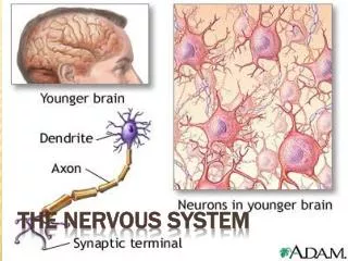
Histology of Nervous Tissue • Two principal cell types • Neuroglia • Neurons (nerve cells)
Neuron Cell Body (Perikaryon or Soma) • Biosynthetic center of neuron • Synthesizes proteins, membranes, and other chemicals • Rough ER (chromatophilic substance or Nissl bodies) • Most active and best developed in body • Spherical nucleus with nucleolus • Some contain pigments • In most, plasma membrane part of receptive region • Most neuron cell bodies in CNS • Nuclei – clusters of neuron cell bodies in CNS • Ganglia – lie along nerves in PNS
Figure 11.4a Structure of a motor neuron. Dendrites (receptive regions) Cell body (biosynthetic center and receptive region) Nucleus Axon (impulse- generating and -conducting region) Myelin sheath gap (node of Ranvier) Nucleolus Impulse direction Chromatophilic substance (rough endoplasmic reticulum) Axon terminals (secretory region) Schwann cell Terminal branches Axon hillock
Figure 11.4b Structure of a motor neuron. Neuron cell body Dendritic spine
Myelin Sheath • Composed of myelin • Segmented sheath around most long or large-diameter axons • Function of myelin • Nonmyelinated fibers conduct impulses more slowly
Table 11.1 Comparison of Structural Classes of Neurons (1 of 3)
Table 11.1 Comparison of Structural Classes of Neurons (2 of 3)
Functional Classification of Neurons • Three types • Sensory (afferent) • Motor (efferent) • Interneurons
Table 11.1 Comparison of Structural Classes of Neurons (3 of 3)
Role of Membrane Ion Channels • Large proteins serve as selective membrane ion channels • Two main types of ion channels • Leakage (nongated) channels • Gated
Role of Membrane Ion Channels:Gated Channels • Three types • Chemically gated (ligand-gated) channels • Voltage-gated channels • Mechanically gated channels
Figure 11.6 Operation of gated channels. Chemically gated ion channels Voltage-gated ion channels Open in response to binding of the appropriate neurotransmitter Open in response to changes in membrane potential Receptor Neurotransmitter chemical attached to receptor Membrane voltage changes Chemical binds Closed Open Closed Open
Resting Membrane Potential:Differences in Ionic Composition • ECF has higher concentration of Na+ than ICF • ICF has higher concentration of K+ than ECF • K+ plays most important role in membrane potential
Action Potentials (AP) • Principle way neurons send signals • Principal means of long-distance neural communication • Occur only in muscle cells and axons of neurons • Brief reversal of membrane potential with a change in voltage of ~100 mV
Figure 11.11 The action potential (AP) is a brief change in membrane potential in a “patch” of membrane that is depolarized by local currents. The big picture The key players Voltage-gated Na+ channels Voltage-gated K+ channels Resting state Depolarization 1 2 Outside cell Outside cell +30 Repolarization 3 3 Inside cell Inactivation gate Inside cell Activation gate 0 Membrane potential (mV) Action potential Closed Opened Inactivated Closed Opened 2 Hyperpolarization 4 The events Threshold –55 Potassium channel Sodium channel –70 1 1 4 0 1 2 3 4 Time (ms) Activation gates Inactivation gate The AP is caused by permeability changes in the plasma membrane: Resting state 1 +30 Action potential 3 0 Membrane potential (mV) Relative membrane permeability Na+ permeability Hyperpolarization Depolarization 2 4 2 K+ permeability –55 –70 1 1 4 0 1 2 3 4 Time (ms) Repolarization 3
Figure 11.11 The action potential (AP) is a brief change in membrane potential in a “patch” ofmembrane that is depolarized by local currents. (1 of 3) 1 Resting state. No ions move through voltage-gated channels. 2 3 Depolarization is caused by Na+ flowing into the cell. Repolarization is caused by K+ flowing out of the cell. +30 3 0 4 Hyperpolarization is caused by K+ continuing to leave the cell. Membrane potential (mV) Action potential 2 Threshold –55 –70 1 1 4 0 1 2 3 4 Time (ms)
Figure 11.11 The action potential (AP) is a brief change in membrane potential in a “patch” ofmembrane that is depolarized by local currents. (1 of 3) 1 Resting state. No ions move through voltage-gated channels. +30 0 Membrane potential (mV) Action potential Threshold –55 –70 1 1 0 1 2 3 4 Time (ms)
Figure 11.11 The action potential (AP) is a brief change in membrane potential in a “patch” ofmembrane that is depolarized by local currents. (1 of 3) 1 Resting state. No ions move through voltage-gated channels. 2 Depolarization is caused by Na+ flowing into the cell. +30 0 Membrane potential (mV) Action potential 2 Threshold –55 –70 1 1 0 1 2 3 4 Time (ms)
Figure 11.11 The action potential (AP) is a brief change in membrane potential in a “patch” ofmembrane that is depolarized by local currents. (1 of 3) 1 Resting state. No ions move through voltage-gated channels. 2 3 Depolarization is caused by Na+ flowing into the cell. Repolarization is caused by K+ flowing out of the cell. +30 3 0 Membrane potential (mV) Action potential 2 Threshold –55 –70 1 1 0 1 2 3 4 Time (ms)
Figure 11.11 The action potential (AP) is a brief change in membrane potential in a “patch” ofmembrane that is depolarized by local currents. (1 of 3) 1 Resting state. No ions move through voltage-gated channels. 2 3 Depolarization is caused by Na+ flowing into the cell. Repolarization is caused by K+ flowing out of the cell. +30 3 0 4 Hyperpolarization is caused by K+ continuing to leave the cell. Membrane potential (mV) Action potential 2 Threshold –55 –70 1 1 4 0 1 2 3 4 Time (ms)
Importance of Myelin Sheaths:Multiple Sclerosis (MS) • Autoimmune disease affecting primarily young adults • Myelin sheaths in CNS destroyed • Treatment • Drugs that modify immune system's activity improve lives • Prevention? • High blood levels of Vitamin D reduce risk of development
The Synapse • Nervous system works because information flows from neuron to neuron • Neurons functionally connected by synapses
Important Terminology • Presynaptic neuron • Postsynaptic neuron
Information Transfer Across Chemical Synapses • AP arrives at axon terminal of presynaptic neuron • Causes voltage-gated Ca2+ channels to open • Ca2+ floods into cell • Synaptotagmin protein binds Ca2+ and promotes fusion of synaptic vesicles with axon membrane • Exocytosis of neurotransmitter into synaptic cleft occurs • Higher impulse frequency more released
Information Transfer Across Chemical Synapses • Neurotransmitter diffuses across synapse • Binds to receptors on postsynaptic neuron • Often chemically gated ion channels • Ion channels are opened • Causes an excitatory or inhibitory event (graded potential) • Neurotransmitter effects terminated
Termination of Neurotransmitter Effects • Within a few milliseconds neurotransmitter effect terminated in one of three ways • Reuptake • By astrocytes or axon terminal • Degradation • By enzymes • Diffusion • Away from synaptic cleft
Neurotransmitters • 50 or more neurotransmitters have been identified • Most neurons make two or more neurotransmitters • Usually released at different stimulation frequencies