Download
1 / 15
150 likes | 243 Vues
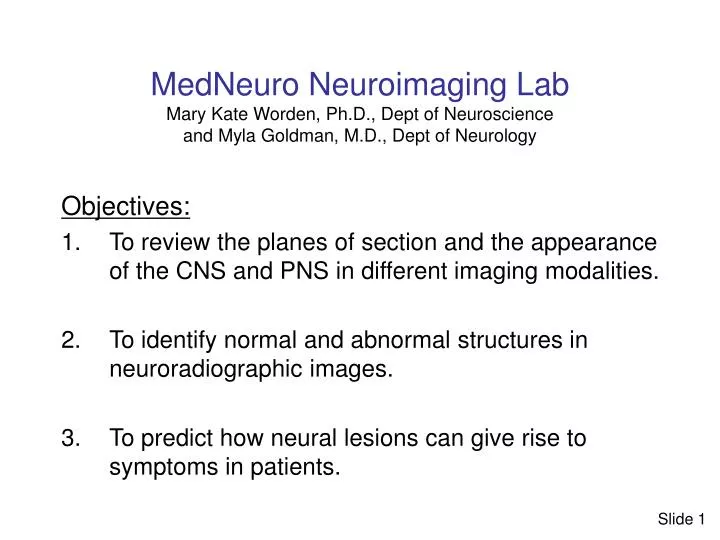
MedNeuro Neuroimaging Lab Mary Kate Worden, Ph.D., Dept of Neuroscience and Myla Goldman, M.D., Dept of Neurology. Objectives: To review the planes of section and the appearance of the CNS and PNS in different imaging modalities.

E N D
MedNeuro Neuroimaging LabMary Kate Worden, Ph.D., Dept of Neuroscience and Myla Goldman, M.D., Dept of Neurology Objectives: • To review the planes of section and the appearance of the CNS and PNS in different imaging modalities. • To identify normal and abnormal structures in neuroradiographic images. • To predict how neural lesions can give rise to symptoms in patients. Slide 1
Q1. Identify the imaging modality, orientation and plane of section for these images. Q2. One of these patients has motor weakness and the other has sensory deficits. Identify the abnormalities in these images and explain the symptoms. A. B. Slide 2
Q3.Identify the internal carotid angiogram and the vertebral artery angiogram. Q4. Identify PCA, ACA, vertebral arteries, internal carotid, superior cerebellar artery, cortical branches of MCA, PCA, lenticulostriate arteries, ophthalmic artery, basilar artery, posterior communicating artery, insular branches of MCA, and PICA. (Hint: at least one artery listed cannot be seen. Why not?) Slide 3
Case study Patient’s lower back A ten year old girl with a history of minimal health care presents with low back pain, increased lower extremity weakness, and incidence of bladder incontinence. Her neural shows decreased function in her dorsal and plantal flexors bilaterally, decreased function of the left quadraceps femoris musculature, decreased reflexes (pateller and ankle) and sensory deficit (decreased sensation to pin prick) of the left L5 dermatome. She also has focal hirsutism of the lower back (indicated by arrow on image). You send her to radiology (next slide). Slide 4
Patient Q5. Identify the imaging modality, the orientation and the plane of section. Q6. Identify the structure indicated by the arrows. Normal Slide 5
Patient L5 Normal Q7. Identify the modality of imaging, the orientiation and the plane of section. What does the arrow point to in the image of the patient? Q8. Identify the level of the conus medullaris in the patient and in the normal image. At what level is a lumbar puncture performed? Slide 6
Neuroradiographs from this patient: . Q9. What do you think caused the abnormalities you see in these images from this patient? Q10. Explain why the clinicians who treated this patient described her spinal cord as “tethered”. Q11. How do these images explain her symptoms? Slide 7
Q12. Identify the imaging modality and plane of section. Q13. Which image is superior to the other? Q14. Identify the lesion and the arterial territory in which it is located. Q15. Identify the cerebral peduncles, the third ventricle and the lateral fissure. Slide 8
Q16. Which of the following functions do you suspect might be impaired in this patient? Explain. sensory (if so, where?) vestibular function motor function (if so, where?) language Slide 9
Authors of a study of schizophrenic patients measured the “gyrification index” in each patient as the ratio of length of the inner cortical contour to the length of the outer cortical contour. Q17. What is the imaging modality and orientation? Q18. Which side of the brain did they measure in these images? Q19. Identify the internal capsule. What is the internal capsule composed of? Slide 10
Authors of a study of schizophrenic patients measured the “gyrification index” in each patient as the ratio of length of the inner cortical contour to the length of the outer cortical contour. Q20. What lobe(s) of the brain are measured in the anterior segment image? In the posterior segment image? Q21. What would you estimate the “gyrification index” to be in these images? Slide 11
The authors of this study reported that schizophrenics have a lower gyrification index than normal patients. Sallet et al (2003) Am J Psychiatry 160:1606-1613, Reduced cortical folding in schizophrenics: An MRI morphometric study Q22. Do you think that the degree of structural abnormality might correlate with the degree of symptoms in these patients? Why or why not? Q23. What do you think caused the the structural abnormalities reported by these authors? Slide 12
Q24. Name the orientation and imaging modality of these sections. Q25. Can you identify structure indicated by the arrows? Slide 13
In the images below the foramen magnum is outlined in red. Q26. Which is normal and which is the patient? How did you decide? Q27. Identify the vertebral arteries in both images. Slide 14
Q28. Identify the following cisterns: cisterna magna, interpeduncular cistern, pontine cistern, chiasmatic cistern, superior cistern. Q29. Name some pathalogical processes that might cause one or more of the cisterns to change shape. Slide 15