Download
1 / 1
10 likes | 212 Vues
0 sec (image 3). 100 sec (image 13). 200 sec (image 23). 300 sec (image 33). CONCLUSION. The results show moderate intraindividual differences but wide variability between individuals.

E N D
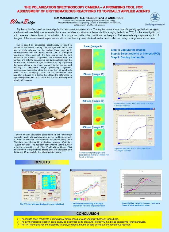
0 sec (image 3) 100 sec (image 13) 200 sec (image 23) 300 sec (image 33) CONCLUSION • The results show moderate intraindividual differences but wide variability between individuals. • The erythemateous reaction could easily be quantified as to area and intensity with a broad capacity to kinetic analysis. • The TiVi technique has the capability to analyze large amounts of data during an erythemateous reaction. THE POLARIZATION SPECTROSCOPY CAMERA – A PROMISING TOOL FOR ASSESSMENT OF ERYTHEMATEOUS REACTIONS TO TOPICALLY APPLIED AGENTS B.M MAGNUSSON1, G.E NILSSON2 and C. ANDERSON1 1Department of Biomedicine and Surgery, Division of Dermatology, 2Department of Biomedical Engineering, Division of Biomedical Instrumentation, Linköping University Hospital, Sweden. Erythema is often used as an end point for percutaneous penetration. The erythemateous reaction of topically applied model agent methyl-nicotinate (MN) was evaluated by a new portable, non-invasive tissue viability imaging technique (TiVi) for the investigation of microvascular tissue blood concentration. In comparison with other traditional techniques, TiVi automatically captures up to 12 images of the microcirculation per minute with a user-friendly computerized system which also can analyze large amounts of data. TiVi is based on polarization spectroscopy of blood in superficial skin tissue. Lineraly polarized light incindent on the skin is partly reflected by the surface layers and partly backsscattered from the dermal tissue. Use of orthogonal polarization filters over both the light source and the light device in the camera suppresses the reflection from the surface, and only the depolarized light backscattered from the dermal matrix reaches the light sensitive array. By separating the colour planes of an image acquired in this manner and applying a dedicated image processing algorithm, spectroscopic information about the amount of red blood cells (RBC) in the underlying tissue can be discovered. The algorithm is based on a theory that utilises the differences in light absorption of RBC and dermal tissue in the red and green wavelength regions. Step 1: Capture the images Step 2: Select regions of interest (ROI) Step 3: Display the results Erythema intensity for a ROI as a function of time in a single individual. Seven healthy volunteers participated in this technology evaluation study. MN solutions were applied to skin occlusively in order to minimise environmental influences using Finn Chambers on Scanpor® application systems (Epicutan, Tuusula, Finland). The application site was the ventral surface of the forearm and the back (20 µl 10 mM MN for 30 sec). TiVi measurement was performed directly after the application and then every 10 seconds for the following 30 minutes. Erythema area for a ROI as a function of time in a single individual. Comparison of photographic and spectroscopic data for 2 selected ROI from 0 to 300 sec. RESULTS Interindividual variablity in seven volunteers (mean of eight application sites). Intraindividual variablity at the eight application sites in a single individual. The TiVi user interface displayed for one individua.l