Download
1 / 51
510 likes | 538 Vues
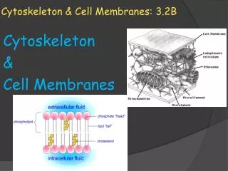
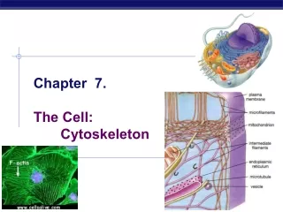
The CELL SURFACE AND CYTOSKELETON 3-D Journey. Chapter 5. The Cell Membrane and Cell Function. Cell Membrane recognizes “self”. Organ rejection Name tags of proteins, carbs and lipids. Cytoskeleton. Cell’s interior scaffolding Protein fibers. Cytoskeleton + cell membrane. Cells framework

E N D
Cell Membrane recognizes “self” • Organ rejection • Name tags of proteins, carbs and lipids
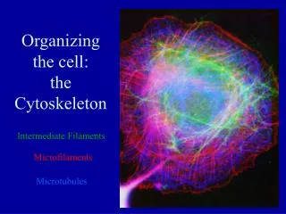
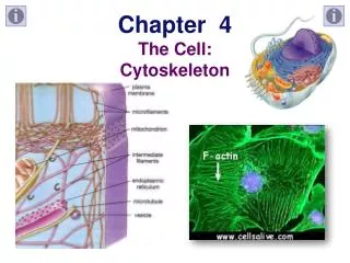
Cytoskeleton • Cell’s interior scaffolding • Protein fibers
Cytoskeleton + cell membrane • Cells framework • Organizes interior of cell • Distinguishes on cell from another
A. Cell Membrane Structure Cell membrane is a phospholipid bilayer embedded with mobile proteins. – Sandwich-like layering repels both charged and uncharged particles Phosphate head of phospholipid is hydrophilic. Fatty acid tails are hydrophobic. Proteins
CM Structure, con’t • Proteins – embedded • Passageway for water soluable molecules and ions • Also act as carriers
CM Structure, con’t • Fluid mosaic – everything moves within the layer
CM Structure, con’t • Receptors – glycoproteins on or within the membrane surface
CM Structure, con’t Ratios of proteins to phospholipids reflect health • Upset = sickness • Multiple sclerosis – not enough fat in cell membranes (cell that are supposed to create mylin • TaySachs – excess lipid in cell membrane (cells that build TOO MUCH mylin Layers of plasma membrane wrapped around an axon
Diffusion: Passive Flow Across a Membrane • General terms: • Aquaeous solution – homogeneous mixture of solute + solvent (water) • Concentration – how much solute is in the solvent
Diffusion, con’t • Diffusion moves substances from high to low concentration • Membranes are selectively permeable • Oxygen, water and carbon dioxide freely cross membranes
Diffusion, con’t • Heat increases the rate of diffusion – kinetic energy is increasing
Diffusion, con’t • Concentration gradient – differences in concentration from low to high or vice-versa • Other types of gradients: • pH • Electrical • Pressure • ions
Diffusion, con’t • Dynamic equilibrium – equal movement back and forth
Osmosis • The diffusion of water • MUST be across a membrane • Controlled by solute concentration inside and outside the cell • Solutes can’t move, so water does
Osmosis, cont • Tonicity • Isotonic-solutes inside and outside have the same concentration
Osmosis, con’t • Hypotonic – hypertonic • Relative terms • Can refer to inside or outside of cell • Hypotonic - solution with the lowersolute concentration. • Hypertonic - solution with the highersolute concentration. • Red blood cell example Pg 70
What is effect of immersing an animalcell in a hypertonic or hypotonic solution?
What is effect of immersing a plant cellin a hypertonic or hypotonic solution? Cell immersed in hypertonic solution Cell immersed in hypotonic solution
Osmosis, con’t • Contractile vacuole – organ that regulates water movement in single cell critter (see video on previous slide) • Turgor pressure – how plant cells regulate how much water can flow into cell – water forced against the cell wall
Transport Proteins • Channels Allow Passive Movement at a faster Rate • Forms an opening (tube) for solutes to pass through • Charge and size regulate what passes • FAST -100 million ions or molecules /second • Form of facilitated diffusion
Transport Proteins, con’t • Carriers move cargo from one side to the other • Carrier protein binds to ion or molecule • Protein changes shape and forces particle to other side of membrane • Can be passive (no ATP- therefore another facilitaed diffusion) or active- uses ATP • Passive facilited 100-1000 per ssecond • Active – can move against the gradient
Transport Proteins, con’t • Pumps use energy to move molecules or ions against a gradient • Sodium potassium pump
Transport Proteins, con’t • Pumps, con’t • Co-transport- see diagram (figure 5.11) pg 72 • Creates gradient • Gradient used to transport substance • Symporter molecule responds to gradient movement
Transport Proteins, con’t • Vesicles perform mass movements by packaging substances • Exocytosis – fluids and large particles out of cell • Ex. Sperm – releases enzymes that penetrate egg
Transport Proteins, con’t • Vesicles, con’t • Endocytosis – capture large molecules and fluid on surface and move into cell • Pinocytosis – just water or solutes and water • Phagocytosis – captures and destroys debris, small organisms, bacteria • Endosome – phagocytic vesicle fuses with a lysosome
Transport Proteins, con’t • Vesicles, con’t • Endocytosis, con’t • Receptor mediated endocytosis • Receptor protein binds to ligand, membrane indents, pulling ligand into the cell • Ex. Liver cells take in cholesterol this way
Transport Proteins, con’t • Trancytosis – combination of endo and exocytosis • Moves particles from one side of the cell to another
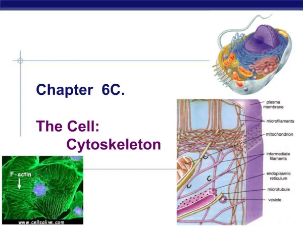
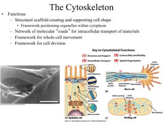
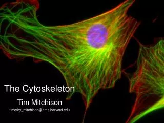
The Cytoskeleton • Tubules and filaments guide organelle movement, provide shape and link molecules
The cytoskeleton, con’t Compare Microtubules and both filaments (figure 5.14 pg 75 aka microfilaments
The cytoskeleton, con’t Microtubules – hollow, thick elements made of the protein tubulin- protein dimmers (pairs) Length adjustable + or – molecules of tubulin Number and arrangement changes based on cell cycle (spindles) Cancer treatments – some effect tubule assembly
The cytoskeleton, con’t Other Functions: • move chromosomes apart during cell division • form cilia & flagella • Cilia- have wave-like motion • 9 microtubule pairs around a central pair (9+2) pattern • Dynein – connects inner and outer proteins (wheel-like) pg 76
The cytoskeleton, con’t • Bad dynein arrangement leads to disease • Clean respiratory track, move egg down fallopian tube, single celled critters swim Leads to Alzheimer’s plaques and tangles
The cytoskeleton, con’t • Flagella • 9+2 arrangement but longer • Whiplike – propulsion • Sperm cells – humans 1, cycad tree - thousands
The cytoskeleton, con’t • Another function – track-way in cell • Moves organelles and proteins within • Squid color changes – rearrange pigment
The cytoskeleton, con’t Microfilaments - long, thin elements made of the protein actin. • Not hollow • Smaller than microtubules Functions: • Strength to cells – withstand stretching and bending) • connect cells to each other • move vesicles & organelles within cytoplasm • Muscle contractions • Actin and myosin interact in sliding filament
The cytoskeleton, con’t Intermediate filaments - elements with diameters in between that of microtubules & microfilaments. Made of various proteins (ie. keratin) Dimers entwined into coiled rods Functions: • maintain cell shape • Inner framework of cell • connect cells to each other & to underlying tissue (skin cells) • Epidermolysis bullosa • Intermediate filaments abnormal – skin blisters easily
Cell Signaling and Response • Cell to Cell communication Intercellular Junctions Structures that connect cells of multicellular organisms to form tissues
Cell Signaling and Response, con’t . Animal cell Connections- types depend on functions Tight Junctions- cell membranes of adjacent cells are fused, creating a tight seal – like a belt Create sheets Ex. cells lining small intestine; cells lining capillaries in brain
Cell signaling and Response, cont • Blood vessels (capillaries) in brain (400 mile-blood brain barrier) • Lipids are chemically soluble – heroin, valium, nicotine, cocaine, alcohol • Oxygen passes • Water soluble – different pathway
Cell Signaling and Response, con’t Desmosomes - intermediate filaments weld cell membranes of adjacent cells together in isolated spots. hold skin cells in place (cell to extrcellular matrix) Ex. skin cells
Cell Signaling and Response, con’t Gap Junctions - channels that link the cytoplasm of adjacent cells, allowing exchange of materials. Ex. heart muscle cells, and muscle cells in digestive track
Cell Signaling and Response, con’t • Cell Walls add structure and allow interactions • Around cells of bacteria, archaea, fungi, algae and plants • Provide shape and volume AND interact
Cell Signaling and Response, con’t • Cell walls, con’t • Composition depends on function of cell, surrounding or life cycle of cell • Plants – mostly cellulose and pectin (like glue)
Cell Signaling and Response, con’t • CW is layers. Oldest layer is most outside layer • Where cell walls meet: middle lamella • Plasmodesmata – link plant cells (tunnels to cells)
Cell Signaling and Response, con’t Cell Adhesion Process that uses membrane proteins called cellular adhesion molecules (CAMs) to direct the migration of cells. Various CAMs function in sequence to: • guide WBCs to injury sites • guide embryonic cells to help form placenta • establish nerve connections involved in learning & memory