Download
1 / 1
10 likes | 131 Vues
2 nd International Meeting on Mammalian Embryogenomics - Paris FRANCE – October 17-20, 2007. A NEW BOVINE EMBRYO SPECIFIC FIBRONECTIN SPLICE VARIANT K. GOOSSENS 1 , A. VAN SOOM 2 , A. VAN ZEVEREN 1 , L.J. PEELMAN 1

E N D
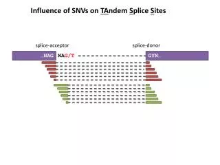
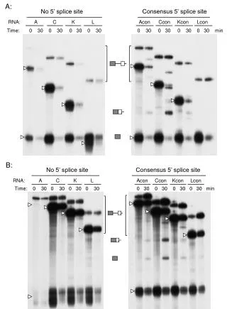
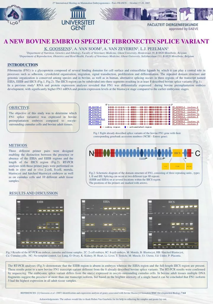
2nd International Meeting onMammalianEmbryogenomics - Paris FRANCE – October17-20, 2007 A NEW BOVINE EMBRYO SPECIFIC FIBRONECTIN SPLICE VARIANT K. GOOSSENS1, A. VAN SOOM2, A. VAN ZEVEREN1, L.J. PEELMAN1 1Department of Nutrition, Genetics and Ethology, Faculty of Veterinary Medicine, GhentUniversity, Heidestraat 19, B-9820 Merelbeke, Belgium 2Department of Reproduction, Obstetrics and Herd Health, Faculty of Veterinary Medicine, Ghent University, Salisburylaan 133, B-9820 Merelbeke, Belgium INTRODUCTION Fibronectin (FN1) is a glycoprotein composed of several binding domains for cell surface and extracellular ligands by which it can play a central role in processes such as adhesion, cytoskeletalorganisation, migration, signal transduction, proliferation and differentiation. The repeated domain structure and genomic organization is conserved among species and in bovine, as well as in human, alternative splicing occurs in three regions of the transcript named EIIIA, EIIIB and IIICS (Fig.1, Fig.2). The IIICS region can be subdivided into three segments resulting in at least 8 described bovine splice variants (Fig.1). In a previous study1 RNA and protein expression analyses revealed that FN1 was differentially expressed during bovine preimplantation embryo development, with significantly higher FN1 mRNA and protein expression levels at the blastocyst stage compared to the earlier embryonic stages. OBJECTIVE The objective of this study was to determine which FN1 splice variant(s) was expressed in bovine preimplantation embryos compared to oocyte-surrounding cumulus cells and bovine adult tissues. N C 1-74 75-286 287-362 IIICS Fig.1 Eightalreadydescribedsplicevariantsof the bovine FN1 gene withtheir correspondinggenebankaccessionnumbers (NCBI – Entrez gene). EIIIB EIIIA Fig.2: Schematic diagram of the domain structure of FN1, consisting of threerepeating units , types I, II and IIII. Splicingcanoccur at two different type III repeats (EIIIB and EIIIA) or at severallocationswithin the IIICS region. The positions of the primers are markedwitharrows. METHODS Three different primer pairs were designed enabling the distinction between the presence or absence of the EIIIA and EIIIB regions and the length of the IIICS region (Fig.2). RT-PCR analyses with those primer pairs were performed on both in vitro and in vivo 2-cell, 8-cell, morula, blastocyst and hatched blastocyst embryos as well as on cumulus cells and 10 different adult tissue samples. EIIIA EIIIB RESULTS AND DISCUSSION In Vitro2C2C 8C 8C M M B B HB HB In Vitro2C2C 8C 8C M M B B HB HB IIICS In Vivo2C 2C 8C 8C B B Cu CuCu NC In Vivo2C 2C 8C 8C B B Cu CuCu NC In Vitro2C2C 8C 8C M M B B HB HB Lu O K H Li T M Ut Ud P Lu O K H Li T M Ut Ud P In Vivo2C 2C 8C 8C B B Cu CuCu NC Lu O K H Li T M Ut Ud P Fig.3 Results of the RT-PCR on embryo, cumulus and tissue samples. 2C: 2-cell embryo, 8C: 8-cell embryo, M: Morula, B: Blastocyst, HB: HatchedBlastocyst, Cu: Cumulus cells, NC: No-templatecontrol, Lu: Lung, O: Ovary, K: Kidney, H: Heart, Li: Liver, T: Testicle, M: Muscle, Ut: Uterus, Ud: Udder, P: Placenta. The RT-PCR analyses (Fig.3) demonstrate that the EIIIB region is absent in embryos whereas the EIIIA region and the full-length IIICS region are present. These results point to a new bovine FN1 transcript variant different from the 8 already described bovine splice variants. The RT-PCR results were confirmed by sequencing. The embryonic splice variant differs from the one(s) expressed in oocyte-surrounding cumulus cells. In bovine adult tissues multiple DNA fragments suggest the presence of more than one transcript isoform, but based on the brighter intensity of a single band it can be concluded that FN1 isoform 5 had the highest expression in all adult tissue samples. REFERENCES: [1] Goossens et al. (2007) Identification and expression analysis of genes associated with bovine blastocyst formation BMC Developmental Biology 7:64 Acknowledgements: The authorswouldlike to thank Ruben Van Gansbekeforhis help in collecting the samples and poster lay-out.