Download
1 / 81
840 likes | 1.73k Vues
Phase Contrast Optics. Abb é Theory. Designed optics for amplitude objects Absorb light without change in phase of light waves Based on assumption of no difference in index of refraction between specimen and background. Criterion for Resolution.

E N D
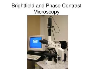
Phase Contrast Optics Theory & Appl. Light Microscopy
Abbé Theory • Designed optics for amplitude objects • Absorb light without change in phase of light waves • Based on assumption of no difference in index of refraction between specimen and background Theory & Appl. Light Microscopy
Criterion for Resolution • Lens must capture undiffracted light plus at least first order of diffracted rays • Combine these in image plane by interference • But — most biological specimens (esp. living) are not amplitude objects • Phase Objects Theory & Appl. Light Microscopy
Phase Objects • Do not absorb light • Difference in index of refraction between specimen and background Theory & Appl. Light Microscopy
Example: Cell • Object 1.25 m thick, i.r. = 1.35; i.r. water = 1.30 (0.05 difference) • Difference in path length for light = 1.25 (0.05) = 0.0625 m • 62.5/500 nm = 1/8 wavelength • /8 = /4 radians = 45° • This is difference in phase of wave passing through cell against wave passing next to cell Theory & Appl. Light Microscopy
Phase Differences • Our eyes cannot see this • Eyes set for amplitude differences, so cell is essentially transparent • But — information is present in light beams from specimen and in image • How do we see this? Theory & Appl. Light Microscopy
Frits Zernike (1888–1966) • Dutch physicist • Developed vector notation for theory of light propagation through phase objects • Invented phase contrast optics in 1930; not manufactured until 1941 by Zeiss Theory & Appl. Light Microscopy
Zernike Phase Vector Diagram For propagation of light through phase object S S = incindent wave P = particle wave P = phase shift of ray through specimen (S = U, undiffracted (0-order) ray P Length of P = amplitude specimen/amplitude medium = transmission ratio Theory & Appl. Light Microscopy
Calculate P by vector addition D U + D = P By the law of sines U P D = of all diffracted orders of light from specimen U = undiffracted light P = resulting specimen light, produced by interference between U and D in image formation Theory & Appl. Light Microscopy
Brightfield Optics • Shifts all vectors in phase equally, and may change all amplitudes equally: U + D = P U = P • No amplitude image • Information in P is present in , not in amplitude — eye cannot see this Theory & Appl. Light Microscopy
Phase Contrast Imaging • Basic principle: • Shift phases (s) and/or amplitudes of U and D differentially • This can produce a change in amplitude of P (length of vector) Theory & Appl. Light Microscopy
In microscope At image plane In specimen D' D' D D P' U U' U' P U' P' Amplitude! U = P
Phase Contrast Optics • Physically separates U and D light and subjects one or the other to phase shift and/or amplitude shift • In theory, any shift of U and D are possible • In practice, a shift of 90° (/4) is appropriate for most biological specimens Theory & Appl. Light Microscopy
Optical Arrangements • Several possible, but major design challenge to keep U and D rays separate and handled differently • In practice, use a hollow cone of light to illuminate specimen • Phase Annulus below condenser • Phase plate at back focal plane of objective • Only 0 order rays from annulus pass through plate Theory & Appl. Light Microscopy
Phase Plate • Rings in phase plate can include • Attenuating layer (absorption but no phase shift), or • Phase-shifting layer (no absorption, phase shift only), or • Any combination of the two Theory & Appl. Light Microscopy
Positive/Negative Phase • Positive Phase Specimen dark against light background (usual now) • Negative Phase Specimen bright against dark background (looks like darkfield optics) Theory & Appl. Light Microscopy
Positive Phase D D' U' U P P' U = P U'> P' Retard D relative to U (move D vector clockwise)
Negative Phase D' D P' U' U P U = P U'< P' Advance D relative to U (move D vector counterclockwise)
Example Systems • Anoptral Phase Contrast Change amplitude of U (soot on ring), no phase shifts for either U or D rays. Bright image — negative phase Popular among algae workers in Great Britain in 50s–60s Theory & Appl. Light Microscopy
Anoptral Phase D No phase shifts on ring D' U U' P P' U = P U'< P' Produces delicate image against brown background
Example Systems • Zernike Phase Contrast Differential changes in amplitude and phase of U and D rays. • All combinations possible: • Amplitude absorption with no phase shift (metal coating) • Phase shift wavefront with no absorption (silica coating) Theory & Appl. Light Microscopy
From: Rose & Pomerat (1960) J. Biophys. Biochem. Cytol. 8:423.
Use/Limitation of Phaseco • Use for qualitative, not quantitative evaluation of specimens • Reasons: • Intensity differences in image not uniquely related to index of refraction differences of specimen • Phase halo— optical artifact Cannot completely separate U and D rays in optics Theory & Appl. Light Microscopy
Intensity Differences • Two points may have same image intensity, but have different values (different i.r.s) • I.e., if IP/IU of at 240° identical to ratio at 320°, then how distinguish different i.r.? Theory & Appl. Light Microscopy
Phase Halo • Serious artifact, most prominent at boundaries of sharp differences in i.r. • Exceeds ability of optics to produce an accurate image • So identification of exact boundary of specimen from image is very difficult Theory & Appl. Light Microscopy
Reducing Phase Halo • Modification of design of phase plate • Apodized Phase Contrast Addition of neutral density filters to phase plate to suppress halo • Optical Process Theory & Appl. Light Microscopy
Reducing Phase Halo • Modification of specimen and medium • Worst halo comes from abrupt i.r. difference between specimen (cell) and medium it is in • Match i.r. of medium to i.r. of specimen to reduce halo • Barer & Joseph (1957) Symp. Soc. Exp. Biol. 10:160–184. • Use of non-osmotic solutes to increase medium index of refraction Theory & Appl. Light Microscopy
Interference Microscopy • Like phaseco in that imaging produces amplitude differences from phase differences in specimen • Quantitative Techniques • Qualitative Techniques Theory & Appl. Light Microscopy
Optical Path Difference • Specimen vs. medium • ' = (s - m)t ' = optical path length t = physical thickness Can measure ', then calculate s = ('/t) + m Theory & Appl. Light Microscopy