Download
1 / 11
110 likes | 119 Vues
Taipei Veterans General Hospital Practices Guidelines Oncology. Oral Cavity Cancer. Version 2010.7. Survival curves of treated oral cancer in Taipei VGH from 2000 to 2004 (N=518). Principles of patient simulation.

E N D
Taipei Veterans General HospitalPractices GuidelinesOncology Oral Cavity Cancer Version 2010.7
Survival curves of treated oral cancer in Taipei VGH from 2000 to 2004 (N=518)
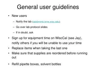
Principles of patient simulation • Patients must have an immobilization device (e.g., aquaplast mask) made prior to treatment planning CT scan. • Shoulder fixation is recommended, esp. with IMRT technique. • The treatment planning CT scan should be performed with IV contrast so that the major vessels of the neck are easily visualized. The treatment planning CT scan must be performed with the immobilization device and in the treatment position. Slice thickness should be at most 0.5 cm.
Principles of Radaition Therapy • Radiation dose • Definitive radiotherapy: 66-74 Gy • Postoperative radiotherapy: 60-66 Gy • Radiation technique: • Intensity-Modulated Radiotherapy (IMRT ) IMRT has been shown to be useful in reducing long-term toxicity in oropharyngeal, paranasal sinus, and nasopharyngeal cancers by reducing the dose to salivary glands, temporal lobes, auditory structures (including cochlea), and optic structures. The application of IMRT to other sites (eg, oral cavity, larynx, hypopharynx, salivary glands) is evolving and may be used at the discretion of treating physicians. • IMRT and Fractionation A number of ways exist to integrate IMRT, target volume dosing, and fractionation. The Simultaneous Integrated Boost (SIB) technique uses differential “dose painting” (66-74 Gy to gross disease; 50-60 Gy to subclinical disease) for each fraction of treatment throughout the entire course of radiation. SIB is commonly used in conventional (5 fractions/week) and the “6 fractions/week accelerated” schedule. The Sequential (SEQ) IMRT technique typically delivers the initial (lower dose) phase (weeks 1-5) followed by the high-dose boost volume phase (weeks 6-7) using 2-3 separate dose plans, and is commonly applied in standard fractionation and hyperfractionation.
Principles of Radiation Therapy • Definitive chemoradiotherapy • Primary and gross adenopathy: • Conventional fractionation: 66-74 Gy (2.0 Gy/fraction; daily) • Altered fractionation: • 6 fractions/week accelerated: 66-74 Gy to gross disease, 44-64 Gy to subclinical disease. • Concomitant boost accelerated RT: 72 Gy/6 weeks (1.8 Gy/fraction, large field; 1.5 Gy boost as second daily fraction during last 12 treatment days) • Hyperfractionation: 81.6 Gy/7 weeks (1.2 Gy/fraction, twice daily) • Neck • Uninvolved nodal stations: 44-64 Gy (1.6-2.0 Gy/fraction)
Principles of Radiation Therapy • Postoperative radiotherapy • Indicated for pT3 or pT4 primary; N2 or N3 nodal disease, selected pT2, N0-N1 disease, nodal disease in levels IV or V, perineural invasion, vascular embolism. • Preferred interval between resection and postoperative RT is 6 weeks. • Primary: 60 Gy (2.0 Gy/fraction) • Neck • Involved nodal stations: 60-66 Gy (2.0 Gy/fraction) • Uninvolved nodal stations: 44-64 Gy (1.6-2.0 Gy/fraction) • Postoperative chemoradiotherapy • Indicated for extracapsular nodal spread and/or positive margins • Consider for other risk features: pT3 or pT4 primary; N2 or N3 nodal disease, nodal disease in levels IV or V, perineural invasion, vascular embolism. • Concurrent chemo with cisplatin + UFUR is commonly used.
Principles of Target volume delineation • Gross Target Volume (GTV) delineation • defined as tumor detected on physical examination or imaging studies. In postoperative cases, the GTV was defined as the preoperative gross tumor volume. • Clinical Target Volume (CTV) delineation • included all potential areas at risk for microscopic tumor involvement by either direct extension or nodal spread. • Including volumes 5 mm around GTV. • Planning Target Volume (PTV) delineation • including a margin for patient motion and setup errors. • 3 to 5 mm margin is usually added to CTV.
Image registration of CT and MRI/PET (if available) should be done for GTV delineation. For oral cavitycancer, adjacent level Ia and Ib of neck are usually included, except for early retromolar trigone tumors (only Ib). Level II to V should be covered for LN (+) cases. Lateral retropharyngealLN (of Rouviere) are rarely involved by oral cavity cancer. (Only few case reports exist). Lateral retropharyngealLN may be included in nasopharyngeal, oraopharyngeal and hypopharyngeal cacner. The following lymph nodes are not included : Level VI: Central compartment group—Lymph nodes in the central compartment of the neck adjacent to the thyroid gland and in the tracheoesophageal groove. Level VII: Superior mediastinal group—Lymph nodes in the superior mediastinum. Contouring guideline
CTV: unilateral or bilateral neck? • For early-stage buccal, gingival, retromolar and hard palate cancer, usually unilateral neck is treated. • For tongue and floor of mouth cancer (midline position), bilateral neck is usually treated. • Locally advanced primary disease, multi-involvement of ipsilateral neck nodes, high pathological grading are associated with contralateral neck lymph node metastasis. (Kurita et al, Oral Oncology 2004; 40:898–903) • For salvage irradiation after local (regional) recurrence, contralateral neck treatment is recommended for patients with ECS. (Liao et al, Ann Surg Oncol 2009; 16:159–170)
RT: Radiation Therapy 3D-CRT: 3D Conformal Radiation Therapy IMRT: Intensity Modulated Radiation Therapy CCRT: Concurrent chemoradiotherapy GTV: Gross Tumor Volume CTV: Clinical Target Volume ITV: Internal Target Volume PTV: Planning Target Volume MRI: Magnetic Resonance Image PET: Positron Emission Tomography Acronyms