Download
1 / 76
800 likes | 1.2k Vues
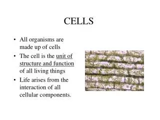
Overview: The Importance of Cells. All organisms are made of cells The cell is the simplest collection of matter that can live Cell structure is correlated to cellular function All cells are related by their descent from earlier cells.

E N D
Overview: The Importance of Cells • All organisms are made of cells • The cell is the simplest collection of matter that can live • Cell structure is correlated to cellular function • All cells are related by their descent from earlier cells
Eukaryotic cells have internal membranes that compartmentalize their functions • The basic structural and functional unit of every organism is one of two types of cells: prokaryotic or eukaryotic • Only organisms of the domains Bacteria and Archaea consist of prokaryotic cells • Protists, fungi, animals, and plants all consist of eukaryotic cells
Comparing Prokaryotic and Eukaryotic Cells • Basic features of all cells: • Plasma membrane • Semifluid substance called the cytosol • Chromosomes (carry genes) • Ribosomes (make proteins)
Prokaryotic cells have no nucleus • In a prokaryotic cell, DNA is in an unbound region called the nucleoid • Prokaryotic cells lack membrane-bound organelles
LE 6-6 Pili Nucleoid Ribosomes Plasma membrane Cellwall Bacterial chromosome Capsule 0.5 µm Flagella A typical rod-shaped bacterium A thin section through the bacterium Bacillus coagulans (TEM)
Eukaryotic cells have DNA in a nucleus that is bounded by a membranous nuclear envelope • Eukaryotic cells have membrane-bound organelles • Eukaryotic cells are generally much larger than prokaryotic cells • The logistics of carrying out cellular metabolism sets limits on the size of cells
LE 6-7 Surface area increases while Total volume remains constant 5 1 1 Total surface area (height x width x number of sides x number of boxes) 750 6 150 Total volume (height x width x length X number of boxes) 125 125 1 Surface-to-volume ratio (surface area volume) 6 6 1.2
The plasma membrane is a selective barrier that allows sufficient passage of oxygen, nutrients, and waste to service the volume of the cell • The general structure of a biological membrane is a double layer of phospholipids
LE 6-8 Outside of cell Carbohydrate side chain Hydrophilic region Inside of cell 0.1 µm Hydrophobic region Hydrophilic region Phospholipid Proteins Structure of the plasma membrane TEM of a plasma membrane
A Panoramic View of the Eukaryotic Cell • A eukaryotic cell has internal membranes that partition the cell into organelles • Plant and animal cells have most of the same organelles
LE 6-9a ENDOPLASMIC RETICULUM (ER Nuclear envelope Flagellum Rough ER Smooth ER NUCLEUS Nucleolus Chromatin Centrosome Plasma membrane CYTOSKELETON Microfilaments Intermediate filaments Microtubules Ribosomes: Microvilli Golgi apparatus Peroxisome Mitochondrion Lysosome In animal cells but not plant cells: Lysosomes Centrioles Flagella (in some plant sperm)
LE 6-9b Nuclear envelope Rough endoplasmic reticulum NUCLEUS Nucleolus Chromatin Smooth endoplasmic reticulum Centrosome Ribosomes (small brown dots) Central vacuole Golgi apparatus Microfilaments Intermediate filaments CYTOSKELETON Microtubules Mitochondrion Peroxisome Chloroplast Plasma membrane Cell wall Plasmodesmata Wall of adjacent cell In plant cells but not animal cells: Chloroplasts Central vacuole and tonoplast Cell wall Plasmodesmata
The eukaryotic cell’s genetic instructions are housed in the nucleus and carried out by the ribosomes • The nucleus contains most of the DNA in a eukaryotic cell • Ribosomes use the information from the DNA to make proteins
The Nucleus: Genetic Library of the Cell • The nucleus contains most of the cell’s genes and is usually the most conspicuous organelle • The nuclear envelope encloses the nucleus, separating it from the cytoplasm
LE 6-10 Nucleus Nucleus 1 µm Nucleolus Chromatin Nuclear envelope: Inner membrane Outer membrane Nuclear pore Pore complex Rough ER Surface of nuclear envelope Ribosome 1 µm 0.25 µm Close-up of nuclear envelope Pore complexes (TEM) Nuclear lamina (TEM)
Ribosomes: Protein Factories in the Cell • Ribosomes are particles made of ribosomal RNA and protein • Ribosomes carry out protein synthesis in two locations: • In the cytosol (free ribosomes) • On the outside of the endoplasmic reticulum (ER) or the nuclear envelope (bound ribosomes)
LE 6-11 Ribosomes ER Cytosol Endoplasmic reticulum (ER) Free ribosomes Bound ribosomes Large subunit Small subunit 0.5 µm TEM showing ER and ribosomes Diagram of a ribosome
The endomembrane system regulates protein traffic and performs metabolic functions in the cell • Components of the endomembrane system: • Nuclear envelope • Endoplasmic reticulum • Golgi apparatus • Lysosomes • Vacuoles • Plasma membrane • These components are either continuous or connected via transfer by vesicles
The Endoplasmic Reticulum: Biosynthetic Factory • The endoplasmic reticulum (ER) accounts for more than half of the total membrane in many eukaryotic cells • The ER membrane is continuous with the nuclear envelope • There are two distinct regions of ER: • Smooth ER, which lacks ribosomes • Rough ER, with ribosomes studding its surface
LE 6-12 Smooth ER Nuclear envelope Rough ER ER lumen Cisternae Ribosomes Transitional ER Transport vesicle 200 nm Rough ER Smooth ER
Functions of Smooth ER • The smooth ER • Synthesizes lipids • Metabolizes carbohydrates • Stores calcium • Detoxifies poison
Functions of Rough ER • The rough ER • Has bound ribosomes • Produces proteins and membranes, which are distributed by transport vesicles • Is a membrane factory for the cell
The Golgi Apparatus: Shipping and Receiving Center • The Golgi apparatus consists of flattened membranous sacs called cisternae • Functions of the Golgi apparatus: • Modifies products of the ER • Manufactures certain macromolecules • Sorts and packages materials into transport vesicles
LE 6-13 Golgi apparatus cis face (“receiving” side of Golgi apparatus) Vesicles coalesce to form new cis Golgi cisternae Vesicles move from ER to Golgi 0.1 µm Vesicles also transport certain proteins back to ER Cisternae Cisternal maturation: Golgi cisternae move in a cis- to-trans direction Vesicles form and leave Golgi, carrying specific proteins to other locations or to the plasma mem- brane for secretion Vesicles transport specific proteins backward to newer Golgi cisternae trans face (“shipping” side of Golgi apparatus) TEM of Golgi apparatus
Lysosomes: Digestive Compartments • A lysosome is a membranous sac of hydrolytic enzymes • Lysosomal enzymes can hydrolyze proteins, fats, polysaccharides, and nucleic acids • Lysosomes also use enzymes to recycle organelles and macromolecules, a process called autophagy Animation: Lysosome Formation
LE 6-14a 1 µm Nucleus Lysosome Lysosome contains active hydrolytic enzymes Hydrolytic enzymes digest food particles Food vacuole fuses with lysosome Digestive enzymes Plasma membrane Lysosome Digestion Food vacuole Phagocytosis: lysosome digesting food
LE 6-14b Lysosome containing two damaged organelles 1 µm Mitochondrion fragment Peroxisome fragment Hydrolytic enzymes digest organelle components Lysosome fuses with vesicle containing damaged organelle Lysosome Digestion Vesicle containing damaged mitochondrion Autophagy: lysosome breaking down damaged organelle
Vacuoles: Diverse Maintenance Compartments • Vesicles and vacuoles (larger versions of vacuoles) are membrane-bound sacs with varied functions • A plant cell or fungal cell may have one or several vacuoles
Food vacuoles are formed by phagocytosis • Contractile vacuoles, found in many freshwater protists, pump excess water out of cells • Central vacuoles, found in many mature plant cells, hold organic compounds and water Video: Paramecium Vacuole
LE 6-15 Central vacuole Cytosol Tonoplast Central vacuole Nucleus Cell wall Chloroplast 5 µm
The Endomembrane System: A Review • The endomembrane system is a complex and dynamic player in the cell’s compartmental organization Animation: Endomembrane System
LE 6-16-3 Nucleus Rough ER Smooth ER Nuclear envelope cis Golgi Transport vesicle Plasma membrane trans Golgi
Mitochondria and chloroplasts change energy from one form to another • Mitochondria are the sites of cellular respiration • Chloroplasts, found only in plants and algae, are the sites of photosynthesis • Mitochondria and chloroplasts are not part of the endomembrane system • Peroxisomes are oxidative organelles
Mitochondria: Chemical Energy Conversion • Mitochondria are in nearly all eukaryotic cells • They have a smooth outer membrane and an inner membrane folded into cristae • The inner membrane creates two compartments: intermembrane space and mitochondrial matrix • Some metabolic steps of cellular respiration are catalyzed in the mitochondrial matrix • Cristae present a large surface area for enzymes that synthesize ATP
LE 6-17 Mitochondrion Intermembrane space Outer membrane Free ribosomes in the mitochondrial matrix Inner membrane Cristae Matrix Mitochondrial DNA 100 nm
Chloroplasts: Capture of Light Energy • The chloroplast is a member of a family of organelles called plastids • Chloroplasts contain the green pigment chlorophyll, as well as enzymes and other molecules that function in photosynthesis • Chloroplasts are found in leaves and other green organs of plants and in algae • Chloroplast structure includes: • Thylakoids, membranous sacs • Stroma, the internal fluid
LE 6-18 Chloroplast Ribosomes Stroma Chloroplast DNA Inner and outer membranes Granum 1 µm Thylakoid
Peroxisomes: Oxidation • Peroxisomes are specialized metabolic compartments bounded by a single membrane • Peroxisomes produce hydrogen peroxide and convert it to water
LE 6-19 Chloroplast Peroxisome Mitochondrion 1 µm
The cytoskeleton is a network of fibers that organizes structures and activities in the cell • The cytoskeleton is a network of fibers extending throughout the cytoplasm • It organizes the cell’s structures and activities, anchoring many organelles • It is composed of three types of molecular structures: • Microtubules • Microfilaments • Intermediate filaments
LE 6-20 Microtubule Microfilaments 0.25 µm
Roles of the Cytoskeleton: Support, Motility, and Regulation • The cytoskeleton helps to support the cell and maintain its shape • It interacts with motor proteins to produce motility • Inside the cell, vesicles can travel along “monorails” provided by the cytoskeleton • Recent evidence suggests that the cytoskeleton may help regulate biochemical activities
LE 6-21a Vesicle ATP Receptor for motor protein Motor protein (ATP powered) Microtubule of cytoskeleton
Components of the Cytoskeleton • Microtubules are the thickest of the three components of the cytoskeleton • Microfilaments, also called actin filaments, are the thinnest components • Intermediate filaments are fibers with diameters in a middle range
Microtubules • Microtubules are hollow rods about 25 nm in diameter and about 200 nm to 25 microns long • Functions of microtubules: • Shaping the cell • Guiding movement of organelles • Separating chromosomes during cell division
Centrosomes and Centrioles • In many cells, microtubules grow out from a centrosome near the nucleus • The centrosome is a “microtubule-organizing center” • In animal cells, the centrosome has a pair of centrioles, each with nine triplets of microtubules arranged in a ring
LE 6-22 Centrosome Microtubule Centrioles 0.25 µm Microtubules Longitudinal section of one centriole Cross section of the other centriole