Download
1 / 37
380 likes | 1.19k Vues
Shoulder Dystocia “Making the Best of a Bad Situation”. Chukwuma I. Onyeije, M.D. Director of Obstetrics and Perinatal Services North Central Bronx Hospital Albert Einstein College of Medicine. Incidence. Varies widely based on criteria used for diagnosis.

E N D
Shoulder Dystocia“Making the Best of a Bad Situation” Chukwuma I. Onyeije, M.D. Director of Obstetrics and Perinatal Services North Central Bronx Hospital Albert Einstein College of Medicine
Incidence • Varies widely based on criteria used for diagnosis. • Gross et al, Toronto General Hospital - 1987 • 0.9 Percent based on coding • 0.2 Percent based on use of maneuvers • Acker et al 1986 • 2 Percent based on assessment of operator • Incidence appears to be increasing as birthweights increase.
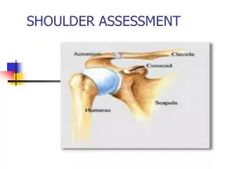
Definition and Diagnosis • “Difficulty encountered in the delivery of the fetal shoulders after delivery of the head.” • Due to impaction of the fetal shoulder behind the symphysis pubis.
ANTEPARTUM FACTORS Maternal Obesity Maternal Diabetes Mellitus Postterm Pregnancy Excessive Weight Gain INTRAPARTUM FACTORS Prolonged Second Stage of Labor Oxytocin Induction Midforceps and Vacuum Extraction Risk Factors Remember, many cases of shoulder dystocia occur with no readily identified risk factors!!!!
Fetal Complications • Fetal Fractures - • In 18 to 25% of cases • Erb’s Palsy - • Although 80% will resolve by 18 months • Perinatal Asphyxia - Uncommon • Neonatal Death - Rare
Maternal Complications • Postpartum Hemorrhage • Vaginal Lacerations • Cervical Lacerations • Puerperal Infection
Management of Shoulder Dystocia • Know the Drill! CALL FOR HELP REMAIN CALM CALL FOR HELP REMAIN CALM Oh, and by the way, don’t forget to call for help.
Management of Shoulder Dystocia • Individuals who MUST be present in the room if shoulder dystocia is anticipated or encountered • Attending physician • Anesthesiologist • Pediatrician • Nursing Staff • “Extra Hands”
Who’s the Boss? • It is important that the conduct of any shoulder dystocia be managed by the most experienced person in the room. • This individual ( generally the attending physician) must have the ability to intervene at any time and should be the only one giving orders.
Preliminary Steps • Call for help and have the team assembled • Drain the bladder • Perform a generous episiotomy • TAKE YOUR TIME, THIS IN AN EMERGENCY, BUT IT IS NOT A RACE!!!
The Principle Maneuvers • Gentle Traction (?) • McRoberts Maneuver • Suprapubic Pressure • Woods’ Corkscrew Maneuver • Delivery of the Posterior Arm
Bilateral Shoulder Dystocia • A bilateral shoulder dystocia. The posterior shoulder is not in the hollow of the pelvis. This presentation oftern requires a cephalic replacement. (C.Pauerstein [ed.], Clinical Obstetrics, Churchill Livingstone, New York, 1987.)
Unilateral Shoulder Dystocia • Unilateral shoulder dystocia is usually easilydealt with by standard techniques. (B. Harris, Shoulder dystocia. Clinical Obstetricsand Gynecology, 1984l 27:106)
Preliminary Measures: • Gentle pressure on the fetal vertex in a dorsal direction will move the posterior fetal shoulder deeper into the maternal pelvic hollow, usually resulting in easy delivery of the anterior shoulder. • Excession angulation (>45 degrees) is to be avoided. (Gabbe, et al., Obstetrics: Normal and Problem Pregnancies, Churchill Livingstone, New York, 1986)
McRobert’s Maneuver • Marked flexion of the maternal thighs unto the abdomen • Decreases the angle of pelvic inclination • Cephalic rotation of the pelvis frees the anterior shoulder
Suprapubic Pressure • Moderate suprapubic pressure is often theonly additional maneuver necessary to disimpactthe anterior fetal shoulder. Stronger pressure canonly be exerted by an assistant. (Gabbe, et al., 1986)
Woods’ Corkscrew Maneuver • Woods' corkscrew maneuver. The shoulders must be rotated utilizing pressure on the scapula and clavicle. • The head is never rotated. (B.Harris, Shoulder dystocia, Clinical Obstetrics and Gynecology, 1984; 27:106.) (B.Harris, Shoulder dystocia, Clinical Obstetrics and Gynecology, 1984; 27:106.)
Woods’ Corkscrew Maneuver • Delivery may be facilitated by counterclockwiserotation of the anterior shoulder to the morefavorable oblique pelvic diameter, or clockwise rotation of the posterior shoulder. • During these maneuvers, expulsive efforts should be stopped and the head is never grasped !!
Delivery of the Posterior Arm • To bring the fetal wrist within reach, exert pressure with the index finger at the antecubital junction. (E. Sandberg. American Journal of Obstetrics and Gynecology, 1985; 152: 481.)
Delivery of the Posterior Arm • Sweep the fetal forearm down over the front of the chest.
Delivery of the Posterior Arm • If less invasive maneuvers fail to affect this impaction, delivery should be facilitated by manipulative delivery of the posterior arm by inserting a hand into the posterior vagina and ventrally rotating the arm at the shoulder with delivery over the perineum.
When All Else Fails... • The Rubin Maneuver • The Chavis Maneuver • The Hibbard Maneuver • Fracture of the Clavicle / Cleidotomy • The Zavanelli Maneuver • Symphysiotomy
The Rubin Maneuver • Step 1: The fetal shoulders are rocked from side to side by applying force to the maternal abdomen. • Step 2: If step one is not successful, push the presenting fetal shoulder toward the chest. This will often cause abduction of both shoulders and create a smaller shoulder to shoulder diameter.
The Chavis Maneuver • Described in 1979. • A “shoulder horn” consisting of a concave blade with a narrow handle is slipped between the symphysis and the impacted anterior shoulder. • This used like a shoe-horn as a lever where the symphysis is the fulcrum.
The Hibbard Maneuver • Release of the anerior shoulder is initiated by firm pressure against the infant's jaw and neck in a posterior and upward direction. An assistant is poised, ready to apply fundal pressure after proper suprapublic pressure • As the anterior shoulder slips free, fundal pressure is applied, and pressure against the neck is shifted slightly toward the rectum.Proper suprapubic pressure is continued.
The Hibbard Maneuver • Continued fundal and suprapublic pressure results in an upward-inward rotation of the newly freed anterior shoulder and a further descent in a position beneath the pubic symphysis.
The Hibbard Maneuver • As a result of the previous maneuvers, the transverse diameter of the shoulders is reduced. • Lateral (upward) flexion of the head releases the posterior shoulder into the hollow of the sacrum.
Fracture of the Clavicle • The anterior clavicle is pressed against the ramis of the pubis. • Care should be taken to avoid puncturing the lung by angling the fracture anteriorly. • Theoretically, a fracture of the clavicle is less serious than a brachial nerve injury and often heals rapidly.
The Zavanelli Maneuver • First described in 1988 • Consists of cephalic replacement and then cesarean delivery. • Mixed reviews in the literature.
... Don’t Even Think About It... • Symphysiotomy is a dangerous procedure with substantial risk to maternal health and well being. • It is difficult to justify this procedure for shoulder dystocia in modern medicine.
Conclusions • Although shoulder dystocia represents a catastrophic event in obstetrics, a well-reasoned plan of action with adequate support and skilled personnel can reduce fetal morbidity. • Proper patient selection and awareness of risk factors for shoulder dystocia can also reduce morbidity.
Although half of shoulder dystocias occur in infants weighing less than 4000 gms…. The incidence of shoulder dystocia is directly related to fetal size.
Complications Associated with Symphysiotomy • Vesicovaginal Fistula • Osteitis Pubis • Retropubic Abscess • Stress Incontinence • Long Term Walking Disability / Pain
Q: Can Cesarean Sections for Suspected Macrosomia Reduce the Rates of Shoulder Dystocia? • Sensitivity of clinical estimates of BW > 4500 gms is only 20% • USG is not very accurate at extremes of EFW • Most cases of shoulder dystocia occur in infants of average weight • The incidence of birth trauma in large infants is not trivial • (2.5% with BW > 4500 gms) A: NO
Top Reasons for Successful Claims Against Obstetricians in Cases of Shoulder Dystocia • Inappropriate obstetrical delivery notes • Absence of delivery notes • Failure to document the dystocia • Failure to document use of McRobert’s maneuver • Lack of prenatal documentation or follow-up of • Abnormal or borderline GTT • Unexpected large maternal weight gain. Harvard Risk Management Foundation (1994) www.rmf.org
Things To Do After Dystocia Occurs • Check for and treat reproductive tract injuries • Pediatric neurology and neonatology consultation • Document a detailed delivery note, including maneuvers used • Explain the occurrence of dystocia to the parents of the infant • Do not finger-point • Be truthful, but avoid discrepancies in notes by doctors, midwives and nurses. Harvard Risk Management Foundation (1994) www.rmf.org