Download
1 / 51
640 likes | 1.63k Vues
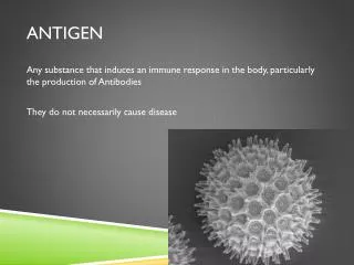
Antigen presenting cells. Antigen presenting cells - (1) dendritic cells (2) macrophages (3) B cells. Morphology of Dendritic Cells. Paradigm: Immunity is the result of co-evolution of microorganisms and the immune system. Gram-. Gram+. Fungi. Parasites. Virus. INFECTIOUS NON SELF.

E N D
Antigen presenting cells • Antigen presenting cells - (1) dendritic cells (2) macrophages (3) B cells
Paradigm: Immunity is the result of co-evolution of microorganisms and the immune system Gram- Gram+ Fungi Parasites Virus INFECTIOUS NON SELF Dendritic cells (DC): the sentinels of the immune system
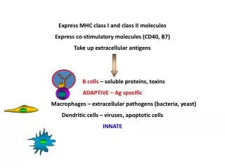
DC orchestrate both innate and adaptive immunity • Phagocytosing microorganisms • Participating to inflammatory responses • Activating appropriate T cell responses
Myeloid precursor Monocytes MOUSE DCs CD8a- and CD8a+ DC Common DC precursor Myeloid- Lymphoid Precursor HSC Langerhans cells Lymphoid precursor ? Plasmacytoid cells Monocytes HUMAN DCs Myeloid precursor CD11b+CD33+ DC Myeloid- Lymphoid Precursor Common DC precursor HSC Langerhans cells Lymphoid precursor Plasmacytoid cells
DCs progenitors are generated in the bone marrow. They give rise to circulating DCs precursors. Circulating DCs precursors enter nonlymphoid tissues as immature DCs. DC are scattered throughout all non lymphoid tissues where they reside in a resting, (so-called immature) state. In the absence of ongoing inflammatory and immune responses, they constantly migrate at low rate to draining lymph nodes. DCs precursors DCs progenitors Immature DCs In inflammatory conditions, immature DC migrate to draining lymph nodes where after maturation (mature DC), they prime the rare circulating naïve antigen-specific lymphocytes. Mature DCs
There are no lineage-specific surface markers that are expressed on all DC. Moreover, DC are a heterogenous cell population. However, it has been clearly demonstrated that at least 3 distinct types of DC can be generated. Mice: myeloid DC CD11b+CD11c+, myeloid DC CD11b+ CD11c+/-, CD4+, lymphoid DC CD11b- CD8a+ The existence of different subsets of DC has lead to possibility that they can perform unique functions.
Plasmacytoid Dendritic Cells • A specialized subpopulation of DC. • Resemble plasma cells in morphology – also express some B cell surface markers (e.g. CD45R [B220]). • In response to some viral infections, pDC synthesize high levels of type I IFNs – cytokines have direct antiviral effects and activate NK cells. • Plasmacytoid DC also are a significant source of IL-12 during the early response – enhance IFN-g production by NK cells, and, subsequently, CD4 and CD8 T cells.
Process of Dendritic Cell Migration to Lymph Nodes
Growth factor-dependent DC conditioned medium GM-CSF or GM-CSF IL-4 bone marrow or peripheral blood Maturation stimulus Immature DC Mature DC Apoptotic DC
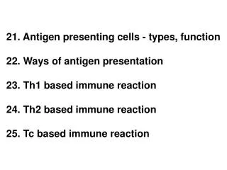
Phagocytosis by DC triggered membrane- ruffling conventional phagocytosis macro pinocytosis forced endocytosis coiling phagocytosis phagosomes macro- pinosomes spacious vacuoles phagosomes phagosomes/ cytosol/replicative vacuoles • Binding of bacteria to the cell surface • Actin polymerization pseudopod extension around the cell surface • Engulpment • Actin depolymerization • Phagosome maturation transport event
The innate repertoire: the specificity of DC recognition is mediated by an extended family of receptors binding a variety of ligands Gram- bacteria Gram+ bacteria Fungi viruses Poli I:C CpG LPS/LTA FcR CR3 (Mac-1) FcR FcR CD11c TLR 5: flagellinR Immature DC ScavengerR TLR 2: LTA/ Gram+ MARCO R TLR 4: LPS/ Gram- TLR 9: CpG CD14 Collectins DEC205 PAMP ‘s : Pathogen-Associated Molecular Patterns conserved structures produced only by microorganisms but not by the host
Toll-like receptors bacterial glycolipids & triacylated bacterial lipopeptides zymosan, peptidoglycan & diacylated bacterial lipopeptides E. coli LPS, hsp60, RSV F protein CpG DNA, chromatin dsRNA Flagellin Imiquimod CD14 ? ? TLR2/1 TLR2/6 TLR3 TLR4 TLR5 TLR7 TLR8 TLR9 TLR10
Toll-like receptor signaling pathways
Gram- Gram+ Fungi Virus Regulated expression of key molecules for the Initiation of adaptive immune responses INFECTIOUS NON-SELF PAMP’s Mature DC Immature DC LPS, LTA, CpG, PGN cytokines Maturation process T cell activation cytokines NK CD8 CD4 Adaptive responses Innate responses
MATURATION PROCESS Migratory activity Cytoskeleton rearrangements MHC and costimulatory molecules upregulation
Migration • Maturing DCs migrate into the T cell area of lymphoid organs. Immature Dcs express a variety of chemokine receptors (CCR1, CCR5, CXCR1 and CCR6) that participate in their recruitment to inflammed tissue and/or to allow their residency into non-lymphoid tissue. Exemple: Imm. Dcs express CCR6 the receptor for MIP-3a that is constitutively express in liver and lungs. Upon maturation, there is a downregulation of receptors for chemokines produced at the site of inflammation and upregulation of CCR7. SLC is a ligand for CCR7 and is expressed at high levels by HEVs in LNs and by stromal cells in T cell areas of many secondary lymphoid organs. ELC (other ligand of CCR7) made in T cell areas of lymphoid tissue. SLC (6Ckine) and ELC (MIP-3b) act together to direct DC migration to T cell areas Of lymphoid tissue and to promote encounter with T cells. Adhesion molecules Banchereau J. et al. Immunobiology of dendritic cells Ann. Rev. Immunol. 18:767-811, 2000.
Immature DC CCR1 CCR2 CCR4 CCR5 CCR6 CXCR1 CXCR4 Mature DC CCR7 MIP-1a, RANTES, MCP-3, MIP-5 MCPs TARC, MDC MIP-1a, MIP-1b, RANTES MIP-3a IL-8 SDF-1 MIP-3b, SLC (6Ckine) Expression of Chemokine Receptors on Dendritic Cells Receptor Ligand Abbreviations: DC, dendritic cell; MIP, macrophage inflammatory protein; RANTES, regulated on activation, normal T cell expressed and secreted; MCP, monocyte chemoattractant protein; TARC, thymus and activation-regulated chemokine; MDC, monophage derived chemokine; SDF, stromal derived factor; IL, interleukin; SLC, secondary lymphoid-tissue chemokine.
Functional downregulation of chemokine receptor expression during DC maturation Control LPS 0.5 h LPS 1h RNAse protection assay Time 0 30’ 1 h 2 h 3 h 24 h LPS - + + + + + CCR1 [Ca2+] : nM L32 CLPS 2 h LPS 3 h LPS 24h GAPDH 20 sec FURA-2 loaded D1 cells stimulated with MIP-1
Developmental Stages of Dendritic Cells Developmental stages of dendritic cells (DCs) in vivo. The generation of DC precursors in the bone marrow, the recruitment of immature DCs in peripheral tissues, and the migration of DCs into the lymphoid organs are illustrated. The maturation of DCs into potent antigen-presenting cells in case of infection or inflammation and their migration have been amply documented (right), but there is also evidence that in the "steady state“ – that is, in the absence of a "danger signal“ – these immature DCs may migrate into the lymphoid organs while remaining at the immature stage (left). The phenotype of the DC migrating in baseline conditions is still unclear. The movement of maturing DCs and the "constitutive" migration of immature DCs have been shown to depend on chemokine gradients.
MATURATION PROCESS Migratory activity Cytoskeleton rearrangements MHC and costimulatory molecules upregulation
control cells MARCO- positive cells
Overview of Dendritic Cell Maturation
MATURATION PROCESS Migratory activity Cytoskeleton rearrangements MHC and costimulatory molecule upregulation
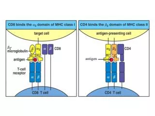
Molecules Involved in T Cell – DC Interactions
Dendritic Cells in Association with T Cells in vivo.
Innate immunity regulates the expression of key molecules for the initiation of the adaptive immune response B71/2 CD40 MHC II MHC l S. thyphimurium E. coli l. monocytogenes M.smegmatis BCG S. aureus S. pyogenes S. pneumococus S. gordonii Lactococco Lactobacillo Mature Immature MHC class I MHC class II B7.1 B7.2 CD40 2.4G2
Peptides bind to MHC molecules through structurally related anchor residues
Peptides that bind MHC class II molecules are variable in lenght
MHC class I loading Antigen Endogenous proteins Exogenous proteins +/- Ubiquitin Proteasome + PA28 Golgi HLA class I synthesis Peptides TAP Calnexin 2m ERp57 Calreticulin Tapasin Endoplasmic reticulum
Cross-Presentation of Antigen by Dendritic Cells • Cross-presentation is the ability of dendritic cells to deliver exogenous antigens to the class I MHC processing and presentation pathway for the activation of CD8 T cells. • The process is rapid, occurring within 3 to 4 hours after antigen uptake. • The process requires a functional endocytic pathway –also requires a functional TAP complex, proteosome function and normal transport from the ER-Golgi to the cell surface. • The process is inhibited if the phagocytic and macropinocytic activities of the DC are inhibited. • Suggests that the DC degrades exogenous antigen in a lysosomal compartment, and then releases relatively long-length peptides into the cytoplasm, where they are ubiquinated, further cleaved by the proteosome, and then delivered to the ER by TAP.
Peptides derived from phagocytosed antigens can be presented to CD8+ T cells on MHC class I. Phenomenon called CROSS-PRESENTATION Soon after or during formation, phagosomes fuse with the ER. After antigen export to the cytosol and degradation by the proteasome, peptides are translocated by TAP into the lumen of the same phagosomes, before loading on phagosomal MHC class I molecule. Therefore, cross-presentation in dendritic cells occurs in a specialized, self-sufficient, ER-phagosome mix compartment.
Dendritic Cells Deliver Exogenous Antigens to the Class I MHC Pathway
The Class II MHC Antigen Processing and Presentation Pathway
Function of HLA-DM (H-2M in Mice)
HLA-DM is Physically Located In the MIIC Vesicle
Phenotypic Changes Associated with Antigen Presentation
Immature DC are less efficient in MLR when compared to mature DC MLR cpm x 10-3 120 mature immature 100 80 60 40 20 0 0,1 1 100 10 APC (x 10-3)
. Allogeneic CD4 T cell proliferation CD4 iDC No DCs mDC 48 h CFSE 72 h FSC
Allogeneic CD8 T cell proliferation CD8 iDC No DCs mDC 48 h CFSE 72 h FSC
Comparison of Antigen Presentation Abilities of DCs and Macrophages
Functional Features of Dendritic Cells Potency Small numbers of DCs pulsed with low doses of antigen stimulate strong T-cell responses. Primary responses Naive and quiescent T cells can be activated with antigens on DCs. Physiology CD4+ T helpers and CD8+ T killers are primed in vivo.