Download
1 / 13
140 likes | 726 Vues
Tuberculous meningitis. Digital Pathology Collection Case 10 2009 Ref. PM243/86. This is a case of disseminated tuberculosis with classical basal tuberculous meningitis and obstructive hydrocephalus. Clinical Data.

E N D
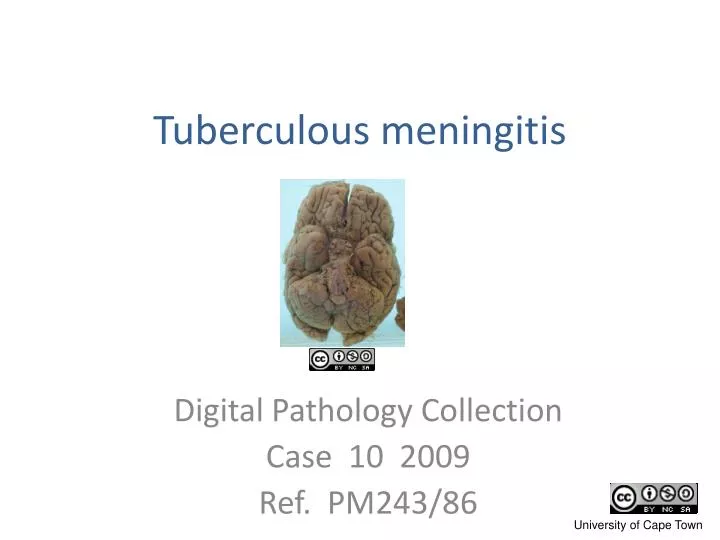
Tuberculous meningitis Digital Pathology Collection Case 10 2009 Ref. PM243/86
This is a case of disseminated tuberculosis with classical basal tuberculous meningitis and obstructive hydrocephalus.
Clinical Data • The subject was a 2 year old coloured boy from poor socio-economic circumstances • He had fever and convulsions and was drowsy and confused. • He also showed drooping of the right side of the mouth and a dragging left leg. • He was thought to have a complicated meningitis, possibly tuberculous.
He then developed acute hydrocephalus. • At surgery very high c.s.f. pressure was noted and an external ventricular drain (“shunt”) was inserted. • He had persistent hyponatraemia, probably due to inappropriate ADH secretion. • He demised two weeks later despite anti-tuberculous and supportive treatment.
The specimen comprises the lower half of the brain and portions of the lungs.
The cut surface of the lung shows segmental consolidation due to extensive tuberculous pneumonia, • as well as enlarged hilar nodes with caseous necrosis.
The brain is swollen; the gyri are flattened. • The meninges are clouded by exudate and there are pinpoint tubercles scattered within them. • The meningitis is most florid at the base of the brain (though we don’t have the vertex for comparison).
The outlet foramina of the fourth ventricle at the base of the brain are occluded by exudate. • Consequently, circulation of c.s.f. out of the internal ventricular system to the subarachnoid space around the brain is obstructed.
The mid-horizontal cut shows the resulting mild hydrocephalus: there is slight dilation of the lateral ventricles. • There is also necrotic disintegration of rostrum of corpus callosum, adjacent gyri and septum pellucidum.
Microscopy showed severe cerebral oedema. • In the meninges were numerous granulomata and a heavy lymphocytic infiltrate. Some necrotising granulomata extended into the cortical substance of the brain, seen macroscopically as tubercles.
Other cases of neurotuberculosis • XII:viii:19 Cerebral tuberculous abscess • link
A selection of cases from the Digital Pathology Collection by the Department of Clinical Laboratory Sciences University of Cape Town is licensed under a Creative Commons Attribution-NonCommercial-ShareAlike 2.5 South Africa Licence The full Digital Pathology Collection is accessible at www.digitalpathology.uct.ac.za If you would like to use an image or other item from our site that is not labelled with the Creative Commons Licence Logo, please contact the curator for permission.