Download
1 / 1
10 likes | 154 Vues
TRANSPALPEBRAL TONOM ETER APPLICATION DURING INTRAOCULAR PRESSURE EVALUATION IN TH E PATIENTS WITH REFRACTION ANOMALY BEFORE AND AFTER KERATOPHOTOREFRACTIVE SURGERY T.B. Dzhafarli, MD., Prof. A.P. Nesterov, MD., A.R. Illarionova, MD. Russian State Medical University, Moscow. B. Purpose :

E N D
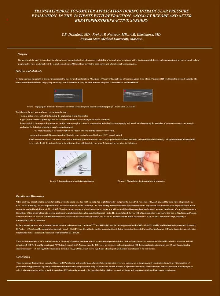
TRANSPALPEBRAL TONOMETERAPPLICATION DURING INTRAOCULAR PRESSURE EVALUATION IN THE PATIENTS WITH REFRACTION ANOMALY BEFORE AND AFTER KERATOPHOTOREFRACTIVE SURGERY T.B. Dzhafarli, MD., Prof. A.P. Nesterov, MD., A.R. Illarionova, MD. Russian State Medical University, Moscow. B Purpose: The purpose of the study is to evaluate the clinical use of transpalpebral scleral tonometry, reliability of its application in patients with refraction anomaly in pre- and postoperational periods, dynamics of eye morphometric rates (pachymetry of the central corneal zone, IOP) and their correlative bond before and after photorefractive surgeries. • Patients and Methods • We have analyzed the results of prospective comparative case series clinical study in 98 patients (194 eyes) with ametropia of various degrees, from which 59 persons (118 eyes) form the group of patients, who had no keratophotorefractive surgery in past history, and 39 patients (76 eyes), who had not been subjected to excimerlaser vision correction. • The following factors were exclusion criteria from the study: • Cornea pathology, potentially influencing the applanation tonometry results; • Upper eyelid and sclera pathology, that are the contraindications for transpalpebral diaton-tonometry • Before and after the surgery all patients were subject to the complete refractive examination, including keratotopography and wavefront-aberrometry. In a number of patients for cornea morphologic evaluation the following procedures have been implemented: • US-biomicroscopy of the corneal optical zone before and two months after laser correction • pachymetry corneal thickness in central (4 points) zone - central corneal thickness (CCT) in each patient • IOP was measured with Goldmann applanation tonometer, pneumotonometer and transpalpebral scleral diaton tonometer using traditional methodology. All ophthalmotone measurements were realized with the patients being in the sitting position with time intervals being 2-3 minutes between two investigators. A B Picture 1 Topographic ultrasonic biomicroscopy of the cornea in optical zone of normal myopia eye (А) and after LASIK (B) Picture1Transpalpebral scleral diaton tonometer Picture 2Methodology for transpalpebral tonometry Results and Discussion While analyzing morphometric parameters in the group of patients who had not been subjected to photorefractive surgeries the mean PCT value was 554.5±32.4 m, and the mean value of applanational IOP – 16.1±2.6 mm Hg, the mean ophthalmotone level evaluated with diaton tonometer – 14.7±2.5 mmHg. At that correlation between values of the applanation tonometer and transpalpebral scleral diaton tonometer was highly reliable (r = 0.73, р±0.005). To define the advantages of scleral tonometry in comparison with the traditional keratoapplanational method we made calculations of real ophthalmotone in the patients of this group taking into account pachymetry, ophthalmometry and applanation tonometry data. The mean value of the real IOP after applanation value conversion was 15.4±2.4 mmHg. Pearson correlation coefficient between real IOP (modified result, received with applanation tonometry) and the value, determined with diaton tonometer was 0.89, р<0.005, which shows high reliability of transpalpebral scleral tonometry. In the groups of patients, who underwent photorefractive vision correction, the mean PCT was 499.8±50.9 m, the mean applanation value IOP – 12.4±2.91 mmHg, modified taking into account keratometry IOP rates – 13.9±3.0 mm Hg, mean diaton-tonometry result – 15.1±2.75 mm Hg. At that we notice approximation of diaton-tonometry figures to the modified applanation IOP value taking into consideration keratometric rates - increase of correlation coefficient from 0.51 to 0.81. The correlation analysis of PCT and IOP results in the group of patients, examined both in preoperational period and after photorefractive vision correction showed reliability of this correlation, p<0.005, reduction of IOP for 1 mm Hg is registered PCT being decreased by 29.7 m. At that, the differences between pre- and postoperational IOP during applanation tonometry was 3.5 mm Hg, and during diaton-tonometry – 1.8 mm Hg, that is statistically dissimilar (t>2, p<0.005), which shows significant advantage of ophthalmotone evaluation if we omit cornea. Conclusion Thus, the cornea thickness is an important factor in IOP evaluation and monitoring, and necessitates the inclusion of corneal pachymetry in the program of examination the patients with suspicion of glaucoma and hypertension, especially after various keratorefractive surgeries while using the traditional corneal methods of ophthalmotonometry. At the same time clinical application of transpalpebral scleral diaton tonometer makes it possible to evaluate IOP using only one device, the procedure being efficient, economical, simple and requires no additional instrument examination.