Download
1 / 36
530 likes | 1.32k Vues
Lumbar Spine. Felix Hernandez, M.D. Some slides and pictures used are with the permission of Dr. Sue Shapiro. Lumbar Spine Bony Anatomy. 5 lumbar vertebrae Massive vertebral bodies with the width greater than the anterior-posterior diameter

E N D
Lumbar Spine Felix Hernandez, M.D. Some slides and pictures used are with the permission of Dr. Sue Shapiro.
Lumbar Spine Bony Anatomy • 5 lumbar vertebrae • Massive vertebral bodies with the width greater than the anterior-posterior diameter • L4 &5 slightly wedge-shaped & anterior convexity (anterior pelvic tilt) these features stabilize and accommodate the increasing body weight supported at the end of spinal column
Facet Joints of Lumbar Spine • Articular process have a distinct concave-convex arrangement: inferior facet sits medial to the superior • superior facet faces medial and posterior and is concave • inferior is convex and faces anterior and laterally- • this limits rotation of the lumbar spine and facilitates flexion, extension and side bending
Lumbar Superior Facets • Superior facet serve as attachment for Multifidus muscle • Multifidus muscle has the same mechanical function as the semispinalis capitis muscle of cervical spine; they are primary extensors of the lumbar and cervical spine
Spinous Process Orientation in Lumbar Spine • Lumbar spine spinous process have horizontal and extended posterior orientation • making it possible to insert a needle b/t the adjacent vertebrae at L3 & L4 for CSF – know as the lumbar puncture
Sacral Spine • Wedged structure made up of 5 fused vertebrae • Superior it articulates with the L5 vertebrae • Inferior with coccyx • Laterally with the ilium
Sacral Spine Anterior /Posterior View • There is a separate anterior and posterior foramina where the sacral nerves exit. The anterior divisions exit from anterior sacral foramen and give rise to the sciatic nerve
L5 S1 Junction • Intervertebral disc located b/t L5 & sacrum is very thick especially along the anterior border • This is where the lumbosacral angle is formed • The most notoriously frequent site for pain in the low back • An increase in lumbar lordosis or increase pelvic tilt results in an increased lumbosacral angle. • Whereas a decreased in lumbosacral angle is due to posterior tilt of pelvis
Lumbar Sacralization • 5th lumbar vertebra may be fused at the lumbosacral joint • The transverse processes forms a pseudoarthrosis at its point of contact with the sacral wing • The L3,4 nerve roots may become irritated on left lateral flexion due to mechanical irritation
Scotty Dog Deformity • Classification of Spondylopathies • Spondylitis- Inflammation of the vertebrae • Spondylosis- Arthritis or osteoarthritis of the vertebrae resulting in pressure being placed on the vertebral nerve root • Spondylolisthesis- Forward slippage of a vertebra on the one below it ( may occur secondary to spondylolysis) • Spondylolysis- Degeneration of a vertebral structure secondary to repetitive stress with no displacement of the vertebral body.
Single Leg Stance Test (Stork Standing Test) • Patient: -Standing with the body weight evenly distributed between the two feet. • Examiner: • -Stand behind your patient ready to support them • patient lifts one leg then places the trunk in hyperextension • Positive Test: • pain is noted in the lumber spine or sacroiliac region of the opposite side of the leg if the lesion is unilateral or both sides of it a bilateral pars fracture. • pain is due to force being placed on the vertebra by the iliopsoas muscle pulling them anteriorly
Sacroiliac Joint • Fusion of first 3 sacral vertebrae and the ilium • Movement of sacrum on the ilium is referred to as Nutation ( flexion) or Counternutation (extension) • Movement of ilium on the sacrum is referred to as torsion – • Anterior torsion occurs when the ASIS moves forward and down ; • posterior torsion occurs when the ASIS moves upward and back
Lumbosacral Trunk • LST is in close approximation to the lumbosacral joint and can cause compression injuries that may cause muscle spasms. • Since L4 & L5 are the principal constituents of the sciatic nerve (L4-S3) any irritation can cause Sciatica
Spinal Nerves and Plexus • There are 31 pairs of spinal nerves • They form networks of nerves or plexus -5 plexuses • Cervical Plexus- C1-C4 • Brachial Plexus-C5- T1 • Lumbar plexus L1-L4 • Sacral plexus L4- S4 • Coccygeal plexus S4-S5
Coccyx • Composed of 3-5 fused vertebral bodies that have no vertebral arch • 1st coccygeal vertebra is the largest and articulates with the sacrum at the sacrococcygeal junction • Serves as a bony origin for muscles of the pelvic diaphragm • Falls on to the coccyx can be very painful and necessitate coccyx excision for pain relief
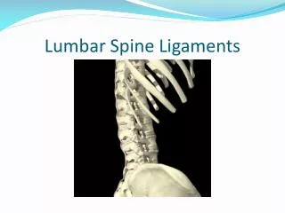
Ligaments of the Lumbar Spine • Anterior Longitudinal Ligament • It’s narrow in the cervical region, increases in width in the lumbar spinal column. • Prevents anterior disc protrusion • Posterior Longitudinal Ligament • it’s widest in the cervical spine but narrows in the thoracic and lumbar spine- • only ½ as wide in lumbar spine as in Cervical spine which is a problem for posterior stability of disc and shifting in the lumbar spine
Ligaments of the Lumbar Spine • Ligamentum flavum • bridges the space b/t adjacent laminae and is highly elastic preventing the chance that lig. Will buckle into the spinal canal • Interspinous Ligament • Broad and thick, resists separation of the spinous processes therefore limiting flexion of the lumbar segments • Supraspinous ligament • most outward ligament that terminates at L4 in 22% of individuals and completely lacking at L5-S1. It limits forward bending of the lumbar spine
LUMBAR DISC • Lumbar disc are significantly thicker and have a greater cross section • Provide 1/3 of the length in the lumbar spine compared to 1/5 in cervical and thoracic spine
Herniated Disc Classifications • Protruded- some eccentric accumulation of the nucleus with slight deformity of annulus • Prolapsed- definite deformity as it works its way through the fiber of annulus • Extruded - nuclear material comes into the spinal canal and runs the risk of impinging adjacent nerve roots • Sequestrated- nuclear material has separated from the disc itself and potentially migrates
Degenerated Disc • Disc herniation can be acute or stress related • Most show signs of previous degeneration • Most common lumbar disc herniation are between L4-L5 and L5-S1 • Next most common herniations are lower two cervical discs
Disc Impingement Symptoms • Intervertebral discs are not innervated thus do not cause pain , it is sensory nerves supplying the ligaments, bony structures, spinal cord or spinal nerves that produce both sensory and motor symptoms • Depending on where herniation takes place the myotome and dermatome patterns associated with the nerve root will cause symptoms
Spinal Stenosis • Defined as a loss of cerebrospinal fluid around the spinal cord due to deformation of the spinal cord, or a narrowing of the neural canal • Common symptom is bilateral leg weakness and numbness with or without sciatica: negative straight leg raise test; + pain on prolonged spine extension exacerbated with ipsilateral trunk lateral flexion
Orthopedic Test for Disc Pathology • Well Straight Leg Raise Test • Milgram Test • Valsalva’s Test
Well Straight Leg Raise Test • The straight leg raise test is an easy test to perform to evaluate the patient for disk herniation. • The patient is placed in the supine position and the well leg is elevated by the clinician up to 70 degrees. • A positive test reproduces radicular pain below the knee along the path of a nerve root in the 30- to 70-degree range of elevation in the leg not being evaluated. • A positive test result can be further verified by lowering the leg 10 degrees from the point of radicular pain and dorsiflexing the foot. • This should produce a similar radicular pain.
Milgram Test • A test which usually confirms pathology either inside or outside the spinal cord sheath. • The test is performed with the patient supine while both legs are held straight out with the heels two to three inches from the table for at least 30 seconds. • The test increases subarachnoid pressure and is positive when the patient is unable to hold the position for 30 seconds without pain • indicating pathology within or outside the spinal cord sheath, such as a herniated disc.
Valsalva Maneuver • The Valsalva maneuver is performed by forcibly exhaling against a closed airway. • Upon the exertion of pressure, neuropathies or radicular pain may be felt, and may indicate impingement on a nerve by an intervertebral disc or other part of the anatomy
Sciatica • Defined as compression and/or inflammation of a spinal nerve making up the sciatic nerve due to a herniated disc, annular tear, myogenic or muscle-related disease, spinal stenosis, facet joint arthropathy, or compression from the piriformis muscle • Typically, if related to herniated disc, radiating leg pain is greater than back pain and increases with sitting and leaning forward , coughing, sneezing and straining • Pain is produced during ipsilateral straight leg raise • With annular tears, back pain is more prevalent and exacerbated with straight leg raise • Differentiate from spinal stenosis because back pain starts usually after walking a limited distance and concomitantly increases as distance increases . • Pain is not reproduced with straight leg raise but can be reproduced with prolonged spine extension and then relived with spine flexion
Orthopedic Test to Evaluate Sciatica • Straight Leg Raise “Lasegue Test” • Kernig/Brudzinski Test
Lasegue Test (Straight Leg Raise Test) • Same straight leg raise test already covered only done on the affected leg with pain being felt in the affected leg.
Kernig/Brudzinski Test • Test Positioning • The subject lies supine with hands cupped behind the head. The examiner stands next to the subject. • Action • The subject is instructed to flex the cervical spine by lifting the head. Each hip is unilaterally flexed to no more than 90 degrees by the subject. The subject then flexes the knee to no more than 90 degrees. The opposite leg remains on the examining table • Positive Finding • The test is confirmed by increased pain (that is either localized or radiates into the lower extremity) with neck and hip flexion. The pain is relieved when the knee is flexed. The pain is indicative of meningeal irritation, nerve root impingement, or dural irritation that is exaggerated by elongating the spinal cord.
Low Back Pain (LBP) • 60 to 80% of the population experiences LBP at some time in their lives • Males and females appear to be equally susceptible • LBP is second only to the common cold as the leading cause of lost work time • Back injuries dominate claims for worker’s compensation • LBP accounts for 10% of all chronic health problems and is ranked 11th among causes for hospitalization in the US • Most cases are idiopathic or unknown origin • Mechanical stress is the primary causal mechanism • Most common among runners , soccer, field hockey , lacrosse, rowers • Mechanism of injury is tight hip flexors, hamstring which produce a forward body lean leading to anterior pelvic tilt and hyperlordosis of the lumbar spine
Lumbar Contusion, Strains, and Sprains • Soft tissue injuries are the most common injuries in the lumbar spine • MOI – Lumbar muscles develop tension to counteract the forward bending moment of the entire trunk when the trunk is in flexion, they are susceptible to strain • Symptoms: Localized pain, increasing with active and resistive motion, radiating pain and neurological deficits