Download

1 / 46
530 likes | 1.08k Vues
CONNECTIVE TISSUE DISORDERS. Presenter: Dr. Gituri Philip Moderator: Dr. Kingori. Outline. Introduction Classification & Types of connective tissue diseases Presentations of CTDs Specific Issues Conclusions. Introduction to CTDs.

E N D
CONNECTIVE TISSUE DISORDERS Presenter: Dr. Gituri Philip Moderator: Dr.Kingori
Outline • Introduction • Classification & Types of connective tissue diseases • Presentations of CTDs • Specific Issues • Conclusions
Introduction to CTDs • May affect predominantly bone, bone and soft tissues, systemic, • May complicate orthopedic procedures • musculoskeletal operations differ in preoperative preparations & outcomes • May be classified into: • Congenital • Acquired • Inflammatory • Immune-mediated
Collagen • 40% of the dry weight of bone is Organic components • Collagen (90% of organic component) • Collagen is primarily type I: provides tensile strength • Type II collagen • 95% of collagen content in articular cartilage • Provides cartilaginous framework and tensile strength • Very stable, with a half-life of approximately 25years
Cont… • Collagen type X • Produced by hypertrophic chondrocytes during enchondralossification: • Growth plate • Fracture callus • Heterotopic ossification formation • Calcifying cartilaginous tumors • Is associated with calcification of cartilage • A genetic defect in type X collagen responsible for • Schmid’smetaphysealchondrodysplasia (affects the hypertrophic physeal zone).
Cont… • Collagen type XI • an adhesive that holds the collagen lattice together
Introduction • Inherited disorders of connective tissue: clinically and genetically diverse group of conditions affecting primarily the skin, joints, and, often, the cardiovascular system. • severity of the musculoskeletal phenotype depends on • the type of mutation • the role & function of the affected protein on musculoskeletal structure
Types of collagen Type Tissues • I Skin, tendon, bone, meniscus, annulus fibrosus • II Articular cartilage, vitreous humor, nucleus pulposus • IIISkin, muscle, blood vessels • IV basement membrane (basal lamina) • V,VI,IX,X articular cartilage • XArticular cartilage, mineralization of cartilage in hypertrophic zone of physis • XI Articular cartilage • XII Tendon • XIII endothelial cells
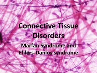
Marfan syndrome • Incidence is 1 in 10,000 • Autosomal dominant; 25% new mutations • Mutation in fibrillin-1 gene on chromosome 15q21; multiple mutations identified • Affected individuals: • Dolichostenomelia • Arachnodactyly • Positive wrist sign (Walker sign) • Positive thumb sign (Steinberg sign)
Arm span-to-height ratio >1.05 • Cardiac defects, especially aortic root dilatation • Scoliosis is seen in 60% to 70% of patients • dural ectasia is common (>60%) • Pectus excavatum and spontaneous pneumothoraces • Pectus carinatum or asymmetric deformity of anterior chest • Superior lens dislocation (ectopia lentis) and myopia • Protrusio acetabuli and severe pes planovalgus
Diagnosis • Clinical assessment • mutational or linkage analysis in familial phenotypes
Classification of Marfans syndrome • Ghent system: 1 major criterion in each of two different organ systems and involvement in a third system • MASS (mitral valve prolapse, aortic root diameter at upper limits of normal, stretch marks, skeletal manifestations of Marfan) phenotype
Treatment • Multi-disciplinary • Nonsurgical • Beta blockers for mitral valve prolapse, aortic dilatation • ii. Bracing for early scoliosis, pesplanovalgus • Surgical • For progressive scoliosis- long scoliosis fusion • progressive protrusioacetabuli, closure of the triradiate cartilage • progressive pes planovalgus, corrective surgery
Ehlers-Danlos syndrome (EDS) • hypermobile joints, hyperextensibleskin, fragile tissues extremely susceptible to trauma • 40% to 50% of patients: mutation in COL5A1 or COL5A2 (type V collagen gene) • 7 types • classic form: AD • Type VI, AR (mutation in lysyl hydroxylase. Severe kyphoscoliosis - characteristic) • Type IV, AD(mutation in COL3A1 thus abnormal collagen III; arterial, intestinal, and uterine rupture seen
Clinical Presentation • Skin: velvety and fragile. Severe scarring with minor trauma common • Joints: hypermobile, esp the shoulders, patellae, and ankles • Pes planus • “double-jointed” fingers • frequent sprains or subluxation of larger joints spontaneously or after slight trauma • 1/3 of patients: aortic root dilatation • vascular subtype: spontaneous visceral or arterial ruptures
c/o chronic joint and limb pain despite normal skeletal radiographs joint hypermobility leads to the onset of OA (3rd or 4th decade) Muscle hypotonia & delayed gross motor development
Importance to surgery • skin splits from trauma, • is relatively painless • does not bleed excessively, • wounds tend to gape • wound margins tend to retract • heal slowly& often become infected • Dehiscence common, & • complete wound breakdown may require repeated suturing or healing by secondary intention
Beighton Criteria for Joint Hypermobility • Passive dorsiflexion of the fifth finger > 90 degrees • Passive apposition of the thumbs to the flexor aspect of the forearm (Beighton sign) • Hyperextension of the elbow > 10 degrees • Hyperextension of the knees > 10 degrees • Ability of the palms to completely touch the floor during forward flexion of the trunk with knees fully extended
Treatment • triad of • anticipatory guidance, • pain management • Physical therapy • Avoid surgery for lax joints; soft-tissue procedures unlikely to work • Progressive scoliosis in type VI (necessary ) • Orthopedicprocedures: Bracing & longer fusions for Progressive scoliosis in type VI
Osteogenesis imperfecta (OI) • Types I through IV : mutation in the COL1A1 and COL1A2 genes • bone that has decreased number of trabeculae and cortical thickness (wormian bone) • Types V through VII no collagen I mutation but • similar phenotype and • abnormal bone on microscopy
Clinical presentation of OI • Child abuse should not be ruled out • types II and III, basilar invagination and severe scoliosis may occur • Olecranon apophyseal avulsion fractures characteristic • dentinogenesis imperfecta, hearing loss, blue sclerae, joint hyperlaxity, and wormian skull bones • frequency of fractures declines sharply after adolescence
Treatment of OI • multi-disciplinary approach. • Manage fractures with light splints • IV & PO Bisphosphonates and growth hormone • severe bowing of the limbs or recurrent fracture: intramedullary fixation is indicated with or without osteotomy. • Progressive scoliosis/basilar invagination is treated with spinal fusion • Transplantation of adult mesenchymal stem cells
Other collagen associated diseases • Scurvy • Acquired: vitamin C deficiency • decrease in chondroitin sulfate and collagen synthesis • greatest deficiency seen in the metaphysis • P/E: microfractures, hemorrhages, and collapse of the metaphysis • Characteristic radiographic findings :line of Frankel and osteopenia of the metaphysis.
Scurvy • Vitamin C (ascorbic acid) deficiency • Produces a decrease in chondroitin sulfate synthesis • defective collagen growth and repair • impaired intracellular hydroxylation of collagen peptides • Clinical features: • Fatigue • Gum bleeding • Ecchymosis • Joint effusions • Iron deficiency • Radiographic findings: • thin cortices & trabeculae and metaphysealclefts (corner sign)
Scurvy cont… • normal bone formation reduced • lacking in tensile strength • defective in structural arrangement • Bow legs • stunted bone growth • swollen joints.
Multiple epiphyseal dysplasia • gene mutation is in COMP • AD • Radiologic findings: irregular, delayed ossification at multiple epiphyses • P/E: Short, stunted metacarpals and metatarsals, • irregular proximal femora, • abnormal ossification (tibial “slant sign” & flattened femoral condyles, patella with double layer) • valgus knees (early osteotomy should be considered), • waddling gait, and early hip arthritis
Treatment of MED • bone survey to differentiate between MED and single epiphyseal dysplasia, as well as to identify all areas of involvement. • Treat limb alignment and perform early joint replacement.
Spondyloepiphyseal dysplasia • Genetic defect: gene encoding type II collagen • abnormal epiphyseal development in the upper and lower extremities • Scoliosis: sharply curved apex over a small number of vertebrae • Retinal detachment and respiratory problems common.
Kniest Syndrome • Defect within type II collagen • AD inheritance • Presentation • short-trunked, disproportionate dwarfism • joint stiffness/contractures, • Scoliosis, kyphosis, • dumbbell-shaped femora, and hypoplastic pelvis and spine • Otitis media and hearing loss frequent • Xray: Osteopenia and dumbbell-shaped bones • Rx: Early therapy for joint contractures. • Reconstructive procedures for early hip degenerative arthritis.
Arthritides Rheumatoid (seropositive) arthritis (RA) • inflammatory autoimmune arthritis • causes joint destruction at a younger age • synoviumthickens, fills with B-cells, T-cells, and macrophages, that erode the cartilage • multiple hot, swollen, morning stiffness. Subcutaneous calcified nodules and iridis • Radiographs: symmetric joint space narrowing, periarticular erosions, and osteopenia
Treatment of RA • Nonsurgical: NSAIDs & DMARDs • Surgical • synovectomy • joint realignment early • joint arthroplasty later stages.
Conclusions • Mutation in the genes coding for various collagen a chains result in a heterogeneous group of heritable conditions (collagenopathies) • Mutations in types II, IX, and XI collagens affects the musculoskeletal, ocular, visual systems, or all three • Diagnosis : clinical findings, radiographic findings, & genetic test results • Follow-up and management: multidisciplinary • Rx is symptomatic and individualized