Download
1 / 26
260 likes | 265 Vues
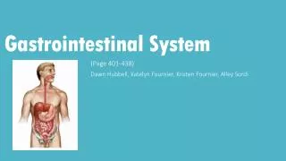
Chapter 3 Abdomen and Gastrointestinal system. By Dr. Amr A. Abd-Elghany. Anatomy and physiology. The abdomen is composed of the abdominal and pelvic cavities and is divided into 9 anatomic regions. It may also be described in terms of quadrants. Abdominal cavity.

E N D
Chapter 3Abdomen and Gastrointestinal system By Dr. Amr A. Abd-Elghany
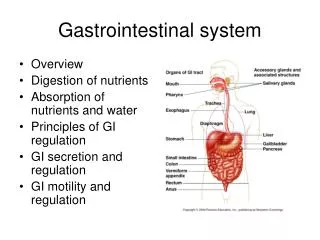
Anatomy and physiology • The abdomen is composed of the abdominal and pelvic cavities and is divided into 9 anatomic regions. • It may also be described in terms of quadrants.
Abdominal cavity Stomach, intestine, hepatobiliary system (liver, gallbladder. Pancrease,) The urinary system, spleen (circulatory system). Pelvic cavity Urinary bladder, portions of the intestines, reproductive organs
Imaging procedures of Alimentary Tract • BARIUM SWALLOW: Detect abnormalities in the upper GIT tract (Esophagus) • BARIUM MEAL: Detect abnormalities in the stomach and duodenum. • BARIUM ENEMA: Detect abnormalities in the lower GIT tract (Colon).
CHOLANGIOGRAPHY • Billiary vessels. • CT/CAT SCAN – • Cross-sectional display of alimentary tract.
Abdominal ultrasonography Radioactive Scan • I.V injection of Radioactive material, then the scanner records uptake of material and produces image. • GastrointestinalEndoscopy • Special tube passed through mouth or anus for visual exam, it can also take biopsy. • CT Guided Biopsy • Percutaneous insertion of needle into organ to remove tissue for microscopic exam. • Magnetic Resonance
Congenital anomalies • Atresia : congenital closure of normal body orifice. • Esophageal Atresia: a discontinuation of the esophagus. • The symptoms are visible soon after birth include: salivation, dyspnea ضيق التنفس. • The radiographic diagnosis: trying to pass radiopaque nasogastric tube as shown in the red arrow.
Malrotation • Aberrations in the normal process of intestinal rotation result in anomalous position of the small and large bowel, with abnormal fixations predisposing the patient to internal herniation and volvulus. • Occurs in an equal male to female ratio.
Inflammatory diseases • Normally the GI system is protected by a mucosal barrier and epithelial cells that remove excess hydrogen ions. • Mucosal blood flow also helps to remove excess acid, thus maintaining a normal pH balance. • Peptic هضمية ulcer is an erosion of the mucous membrane of the lower end of the esophagus, stomach, or duodenum. • The most likely site is the duodenal bulb and the lesser curvature of the stomach. • Duodenal ulcers are found in all ages. • Causes: Non-steroidal anti-inflammatory drugs and Helicobacter pylori. • Symptoms: pain, sometimes bleeding in advanced level carcinoma.
Peptic ulcer • Treatment: • Acid blockage. • Proton inhibitors. • Antibiotics.
Degenerative disease Pharyngitis/Tonsilitis: Is inflammation of Pharynx/Tonsils. Usually due to Streptococcal or viral infection. Oesophagitis: Common cause is reflux of acid contents from stomach called peptic or reflux Oesophagitis (prolonged bed rest). Hernia: • Is abnormal protrusion of viscera through an aperture. • It leads to obstruction or necrosis of the organ.
Diverticular diseases • Diverticula: Is a sac or pouch extending from a hollow organ. Congenital oesophagealDiverticula occur rarely.
Varices Is abnormal dilatation of veins, usually due to obstruction to venous return. Esophageal varices occur due to increased portal blood pressure in liver cirrhosis. This may bleed producing: Severe hematemesis (blood vomiting). Rupture of esophagus.
Disorders of the intestine 1- Obstructions are usually caused by scarring and adhesions, Neoplasm’s, Volvulus. 2- Malabsorption the causes are: A. surgical resection. B. Blind loop syndrome (small Intestinal bacterial overgrowth). C. Lymphatic obstruction. D. Coeliac disease (autoimmune disorder attack intestine villi).
3. Inflammations & Infections • Dysentery: Acute bacterial infection, painful bloody diarrhea. Usually by shigella group. • Salmonella food poisoning: Caused by Salmonella infection. • Typhoid Fever: Systemic infection by Salmonella typhus, causes ulceration of (lymphoid nodules) in intestine. • Tuberculosis of Intestine: Chronic ulceration, fibrosis, stricture with obstruction & perforation.
Ulcerative colitis • is a form of inflammatory bowel disease (IBD). • Ulcerative colitis is a form of colitis, a disease of the colon, that includes ulcers. • The main symptom of active disease is usually constant diarrhea mixed with blood, of gradual onset.
Colon cancer • Colon cancer arises after 40 years. • Adenocarcinoma (mucous secreting glands) is the most common type of colorectal cancer and is derived from the glandular epithelium. • It begins benign adenoma that undergoes a slow malignant transformation through 7 years. • Adenocarcinoma infiltrate the colon wall. • Lesions tend to penetrate and extend into surrounding tissues without causing obstruction.
Common Disorders of Liver & gall bladder are: 1.Cirrhosis –Advanced liver disease results from replacement of liver tissue by fibrosis (scars) and regenerative nodules (lumps that occur due to attempted repair of damaged tissue). • These changes lead to loss of liver function. • Cirrhosis is most commonly caused by alcoholism, hepatitis B and hepatitis C, and fatty liver disease, but has many other possible causes. Some cases are idiopathic (of unknown cause). • Ascites (fluid retention in the abdominal cavity) is the most common complication of cirrhosis. It is associated with a poor quality of life, increased risk of infection, and a poor long-term outcome.
Other potentially life-threatening complications are hepatic encephalopathy (confusion and coma) and bleeding from esophageal varices. • Cirrhosis is irreversible, and treatment usually focuses on preventing progression and complications. In advanced stages of cirrhosis the only option is a liver transplant.
• Cholecystitis - Inflammation of Gall bladder. Jaundice: Yellow discoloration of skin & sclera due to hyperbilirubinemia (increased serum Bilirubin). • Two major types Conjugated or Obstructive & unconjugated or non-Obstructive jaundice. • Jaundice due to biliary obstruction (stone, tumors) is usually conjugated type. • Jaundice in hepatitis is combined type with both conjugated & unconjugated forms due to cell necrosis and obstruction due to inflammation.
Viral Hepatitis: • is liver inflammation due to a viral infection. • It may present in acute (recent infection, relatively rapid onset) or chronic forms. • The most common causes of viral hepatitis are the five unrelated hepatotropic viruses Hepatitis A, Hepatitis B,Hepatitis C, Hepatitis D, and Hepatitis E. • Type B, C & D spread by serum (body fluids, injections, sexual contact) hence they are also known as serum hepatitis. • They cause both acute & chronic recurrent hepatitis. • May complicate with chronic liver failure, necrosis, cirrhosis, and hepatocellular carcinoma. • Vaccine is now available for Hepatitis A, B & C
Under higher-power magnification, ground-glass cells may be visible in chronic HBV infection. • Ground-glass cells are present in 50% to 75% of livers with chronic HBV infection. • Immunohistochemical staining is positive for HBsAg.
Tumors of Liver: • Common tumors of livers are metastasis tumors from abdominal malignancies. • Primary Malignant tumor is hepatocellular carcinoma. • Usually arises over post hepatitis and cirrhosis. • Primary tumors are very aggressive tumor spreads all over body and has poor prognosis. • Benign Tumors are rare.