Download
1 / 20
200 likes | 204 Vues
Chapter 13, part 1. The Spinal Cord and Spinal Nerves. Learning Objectives. Discuss the structure and functions of the spinal cord. Describe the three meningeal layers that surround the CNS.

E N D
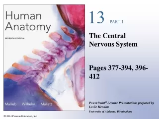
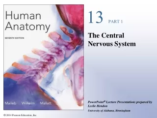
Chapter 13, part 1 The Spinal Cord and Spinal Nerves
Learning Objectives • Discuss the structure and functions of the spinal cord. • Describe the three meningeal layers that surround the CNS. • Describe the major components of a spinal nerve and relate their distribution to their regions of innervation. • Discuss the significance of neuronal pools. • Describe the steps in a neural reflex. • Explain how reflexes interact to produce complicated behaviors.
Divisions of the Nervous System • CNS • Brain and spinal cord • In the white matter, axons arranged in tracts and columns • PNS • Remainder of nervous tissue
Figure 13.1 An Introduction to the Anatomical Organization of the Nervous System Figure 13.1
Adult spinal cord • Localized enlargements provide innervation to limbs • 31 segments • each segment has a pair of dorsal roots and a pair of ventral roots • Filum terminale • Conus medularis • Spinal nerves extend off cord • Mixed nerves
Figure 13.3 Gross Anatomy of the Adult Spinal Cord Figure 13.3
Spinal meninges • Provide physical stability and shock absorption • Three layers • Dura mater • Arachnoid • Pia mater
Dura mater • Covers spinal cord • Tapers to coccygeal ligament • Epidural space separates dura mater from walls of vertebral canal
Figure 13.4 The Spinal Cord and Spinal Meninges Figure 13.4a
Figure 13.4 The Spinal Cord and Spinal Meninges Figure 13.4b
Arachnoid • Interior to dura mater are the subdural space, the arachnoid and the subarachnoid space • Subarachnoid space contains CSF
Pia mater • Meshwork of elastin and collagen fibers • Innermost meningeal layer • Denticulate ligaments extend from pia mater to dura mater
Figure 13.6 The Cervical Spinal Cord Figure 13.6
Sectional anatomy of the spinal cord • White matter is myelinated and unmyelinated axons • Gray matter is cell bodies, unmyelinated axons and neuroglia • Projections of gray matter toward outer surface of cord are horns
Figure 13.7 The Sectional Organization of the Spinal Cord Figure 13.7a
Figure 13.7 The Sectional Organization of the Spinal Cord Figure 13.7b
Horns of spinal cord • Posterior gray horn contains somatic and visceral sensory nuclei • Anterior gray horns deal with somatic motor control • Lateral gray horns contain visceral motor neurons • Gray commissures contain axons that cross from one side to the other
White matter • Divided into six columns (funiculi) containing tracts • Ascending tracts relay information from the spinal cord to the brain • Descending tracts carry information from the brain to the spinal cord PLAY Animation: Spinal cord dissections