Download
1 / 26
270 likes | 284 Vues
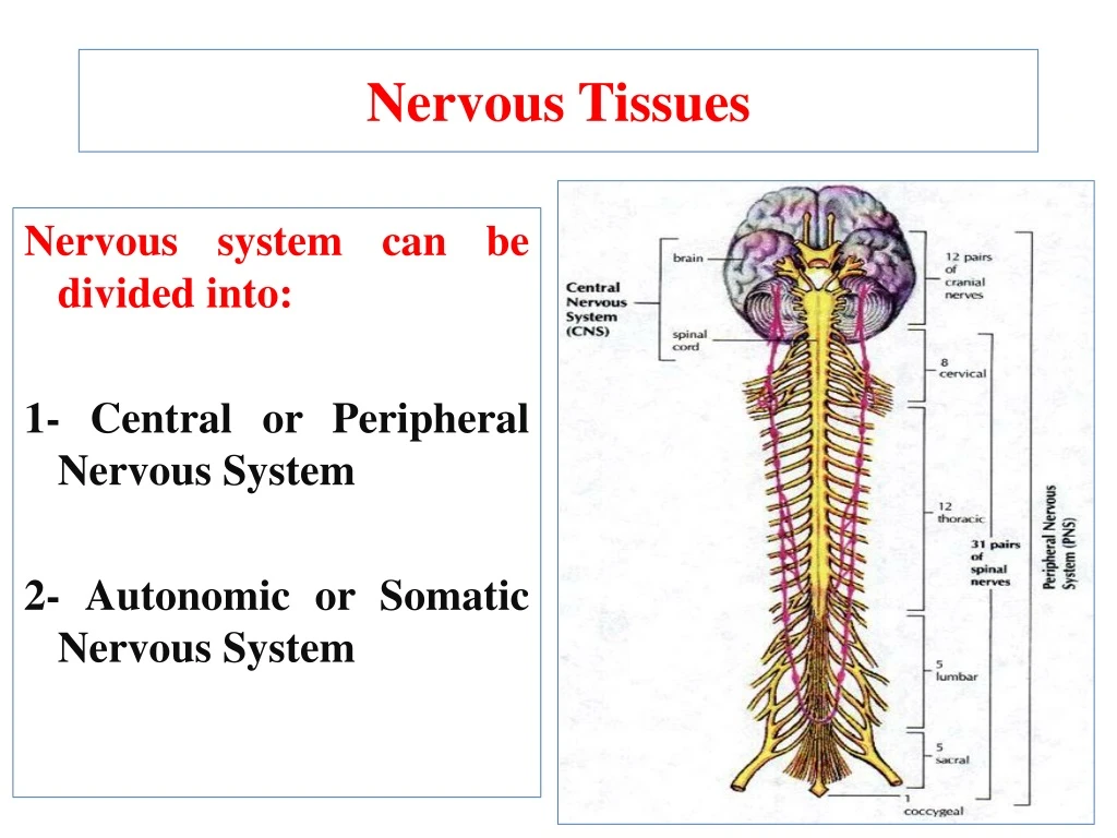
Nervous Tissues. Nervous system can be divided into: 1- Central or Peripheral Nervous System 2- Autonomic or Somatic Nervous System. The central nervous system (CNS) includes the brain and spinal cord.

E N D
Nervous Tissues Nervous system can be divided into: 1- Central or Peripheral Nervous System 2- Autonomic or Somatic Nervous System
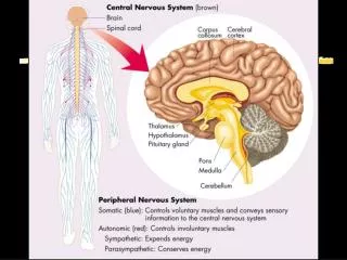
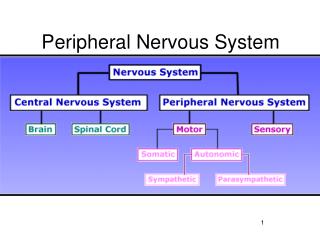
The central nervous system (CNS) includes the brain and spinal cord. • The peripheral nervous system (PNS) includes all other nerve tissues, which could be motor or sensory . • Autonomic NS has both motor and sensory pathways; that controls involuntary visceral functions. • The somatic NS includes all nerve tissues except ANS.
Types of Cells within CNS: • Cells in nervous tissues are of two types: • Neurons (nerve cells) thattransmits impulses. • Neuroglia orSupporting cells. • Little extracellular matrix.
Neurons (Nerve Cells) • Neurons are the functional and structural units of nervous tissue. • Neurons are terminally differentiated cells that are mitotically inactive, i.e. can not divide. • They have conducting pathways, and act as site of integration and analysis of nerve impulses. • Each neuron has a large cell-body (soma) , one axon and several dendrites.
The cell body (soma) could be spherical, ovoid or angular, with a diameter of 5 -150μm. • The cytoplasm contains basophilic granules called Nissl bodies (free ribosomes, and polyribosomes). • Neurotubules: aremicro-tubules and bundles of neuro-filaments found throughout the soma and extend into the axon and dendrites.
Dendrites: • Each neuron has a variable number of branched cytoplasmic processes (dendrites). • They collect incoming messages and carry them toward the cell body then to the axon. • Dendrites may contain few number of mitochondria and Nissl bodies.
Axon: is a single complex cell process that carries impulses away from the soma. • Axon is divided into several regions: • The AxonHillock, the part of the soma leading into the axon, it lacks Nissl bodies. • The axonproper is the main trunk of the axon and tend to have a constant diameter along its length. • Axons of most neurons have a myelin sheath formed by supporting cells (Schwann cells) and interrupted by gaps called nodes of Ranvier. • Myelinated axon segments between the gaps are called internodes.
Types of neurons • Neurons can be classified according to their function into motor, sensory or integrative. • Neurons can be classified according to their axon and dendrites with respect to the shape of cell body into:- • Unipolar neuron: • True Unipolar neuron: in spinal nucleus of trigeminal nerve. • Pseudo-unipolar neuron: primary sensory neurons; single dendrite and axon arise from common stem formed by fusion; present in spinal ganglia.
Bipolar neuron: single dendrite arises opposite origin of axon; receptor neurons for sensation and present in olfactory mucosa, retina and inner ear. • Multipolar neuron: most common and most are motor; several dendrites project from cell body; which are divided into:- • Purkinje cells: the cerebellum. • Polygonal cells: anterior horn cells of spinal cord. • Pyramidal cells: the cerebral cortex.
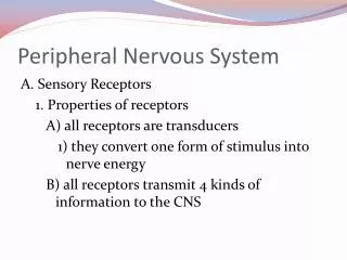
Peripheral nerves (PN) - PN could be afferent (sensory) fibers enter the spinal cord via the dorsal roots or efferent (motor) fibers leave the spinal cord via the ventral roots. - One nerve fiber consists of an axon and its nerve sheath. - Each axon is surrounded by a sheath of Schwann cells.
An individual Schwann cell may surround the axon for several hundred micrometers, and it may, in the case of unmyelinated nerve fibers, surround up to 30 separate axons. • In case of myelinated nerve fibers, Schwann cells form a sheath around one axon and surround this axon with several double layers (up to 100) of cell membrane.
Connective tissue covering nerve fibers - The entire nerve is surrounded by a thick layer of dense connective tissue, the epineurium. - Nerve fibers are frequently grouped into bundles; each bundle is surrounded by a connective tissue layer called perineurium. - The space between individual nerve fibers is filled by loose connective tissue, the endoneurium, which contain fibrocytes, mast cells and macrophages.
Neuroglia (Supportingcells) • Neuroglia cells outside the CNS:- • Schwann cells are the supporting cells of the peripheral nerves. • Satellite cells are specialized Schwann cells in the spinal ganglia, where they form a one-cell-thick covering over the nerve cell bodies (ganglion cells).
2- Neuroglia cells inside the CNS:- • Oligodendrocytes form myelin sheath around axons in the CNS. • Astrocytes (or astroglia) are star-shaped cells, their processes are in contact with a blood vessel (perivascular foot processes). • Microglia is a small cell with a complex shape. Microglia are derived from the same cell line of monocytes, i.e. macrophage precursors.
Ganglia • Ganglia are collection of nerve cells (ganglion cells) outside the CNS, and are of two types: • Spinal ganglia: are surrounded by a connective tissue capsule. • The ganglion cells are pseudounipolar cells, surrounded by layer of flattened satellite cells, present in groups, and separated by fibrous bundles.
Autonomic ganglia: the ganglion cells are multipolar, have dendrites but not surrounded by satellite cells. • Cells are scattered through the ganglia, with few fibers in-between. • Some autonomic ganglia are embedded within the walls of the organs which they innervate (e.g. GIT and bladder).