Download
1 / 1
10 likes | 76 Vues
M3.10. Polyunsaturated fatty acid synthesis de novo is required for calcium release in vascular smooth muscle. N.A.Irvine 1 , C.M.Sibbons 1 , L.R.Grenfell 1 , K.A.Lilycrop 2 , M.A.Hanson 1 and G.C. Burdge 1.

E N D
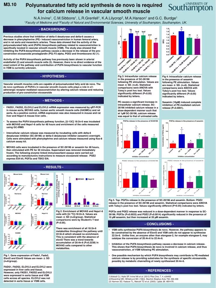
M3.10 Polyunsaturated fatty acid synthesis de novo is required for calcium release in vascular smooth muscle N.A.Irvine1, C.M.Sibbons1, L.R.Grenfell1, K.A.Lilycrop2, M.A.Hanson1 and G.C. Burdge1 1Faculty of Medicine and 2Faculty of Natural and Environmental Sciences, University of Southampton, Southampton, UK. - BACKGROUND - Previous studies show that inhibition of delta-5 desaturase and delta-6 causes a decrease in phenylepherine (PE)- induced vasoconstriction in human femoral artery, and in rat aorta and mesenteric arteries. These data showed that the activity of the polyunsaturated fatty acid (PUFA) biosynthesis pathway related to vasoconstriction is specifically located in vascular smooth muscle (VSM). The study also showed that inhibiting the PUFA-biosynthesis pathway causes a decrease in the release of the pro-constriction eicosanoids prostaglandin (PG) F2 alpha, PGE2 and thromboxane A2 (1). Activity of the PUFA biosynthesis pathway has previously been shown in arterial endothelial (2) and smooth muscle cells (3). However, there is no direct evidence of the exact extent of the pathway and contribution of PUFA biosynthesis to vascular function in VSM is currently unknown. - HYPOTHESIS - Fig 3: Intracellular calcium release in the presence of SC-26196 following PE stimulation. Values are mean ± SD. (n=6). Statistical comparisons were ANOVA with Tukey’s post hoc test. Values significantly different (P<0.05) indicated by letters. PE causes a significant increase in intracellular calcium release. SC-26196 inhibited calcium release in a dose-dependent manner such that at 1µM SC-26196, calcium release was equal to that of untreated cells. Fig 4: Intracellular calcium release in the presence of sesamin following PE stimulation. Values are mean ± SD. (n=6). Statistical comparisons were ANOVA with Tukey’s post hoc test. Values significantly different (P<0.05) indicated by letters. Sesamin (10µM) induced complete inhibition of PE-mediated calcium release (P<0.0001). Vascular smooth muscles cells are capable of polyunsaturated fatty acid de novo. The de novo synthesis of PUFA’s in vascular smooth muscle cells plays a role in α1-adrenergic receptor mediated vasoconstriction by altering calcium release and reducing production of pro-constriction eicosanoids - METHODS - • FADS1, FADS2, ELOVL2 and ELOVL5 mRNA expression was measured by qRT-PCR in mouse aorta, MOVAS cells, human aortic smooth muscle cells (HASMCs) and rat aorta. As a positive control, mRNA expression was also measured in mouse and rat liver and Hepa1-6 mouse liver cells . • To assess the PUFA biosynthesis pathway function, [U-13C] 18:2n-6 was incubated with MOVAS and Hepa1-6 cells for 48 hours and enrichment of the cells measured using GC-IRMS • Intercellular calcium release was measured by incubating cells with delta-6 desaturase inhibitor (SC-26196) or delta-5 desaturase inhibitor (sesamin) overnight. Cells were stimulated with phenylephrine and calcium release measured using fluo-8 calcium assay kit. • MOVAS cells were incubated in the presence of SC-26196 or sesamin for 48 hours, then stimulated with PE for 30 minutes. Supernatant was removed immediately frozen. The following enzyme linked immunosorbent assays were carried out according to manufacturers instructions to measure eicosanoid release: PGE2 express EIA kit, PGF2α and TBX2 EIA. - RESULTS - Fig 5. Top: PGF2α release in the presence of SC-26196 and sesamin. Bottom: PGE2 release in the presence of SC-26196 and sesamin. Statistical comparisons were ANOVA with Tukey’s post hoc test. Values significantly different (P<0.05) indicated by letters. PGF2α and PGE2 release was reduced in a dose-dependent manner in response to SC-26196. PGF2α (P=0.0033) and PGE2 (P=0.0014) significantly reduced in the presence of 10 µM sesamin, but then increased at 20 µM sesamin. Fig 2: Enrichment of MOVAS and Hepa1-6 cells with [U-13C]-18:2n-6. Values are mean ± SD (n=6/group). Statistical comparisons were by Student’s T test (P<0.05). There was enrichment of all 18:2n-6 metabolites throughout the pathway until 22:5n-6 which showed no enrichment. This is consistent with the absence of elovl2 There was a trend towards accumulation of 20:4n-6 (P=0.2238) in MOVAS cells compared to other metabolites. - CONCLUSIONS - • VSM cells synthesise PUFA biosynthesis de novo. However, the pathway appears to be constrained by the absence of Elovl2 and VSM cells do not appear to synthesise 22:5n-6. Unlike liver, an enzyme other than elongase-2, for example elongase-5, may catalyse the conversion of 20:4n-6 to 22:4n-6. • Inhibition of the PUFA biosynthesis pathway causes a decrease in calcium release. This shows that PUFA biosynthesis de novo is involved in calcium release, and thus vasoconstriction, of VSM following PE stimulation. • One possible mechanism by which PUFA biosynthesis may contribute to PE-mediated calcium release is by providing substrates for the synthesis of specific eicosanoids, possibly by metabolic channelling of newly synthesised 20:4n-6. Fig 1. Gene expression of Fads1, Fads2, Elovl2 and Elovl5 Values are mean ± SD (n=6/group). FADS1, FADS2, ELOVL5 and ELOVL2 were expressed in liver cells and tissue. However, only FADS1, FADS2 and ELOVL5 were expressed in aorta tissue and VSM cells across all species. ELOVL2 was not detected in aorta tissue or VSM cells. - REFERENCES - (1)Kelsall CJ, Hoile SP, Irvine NA et al. (2012) Plos One, 7, 4, e34492 (2)Rosenthal MD, Whitehurst MC (1983). Biochim Biophys Acta 750: 490-496 (3) Harmon SD, Kaduce TL, Manuel TD et al. (2003). Lipids 38: 469-476