Download
1 / 6
120 likes | 603 Vues
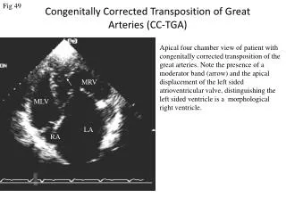
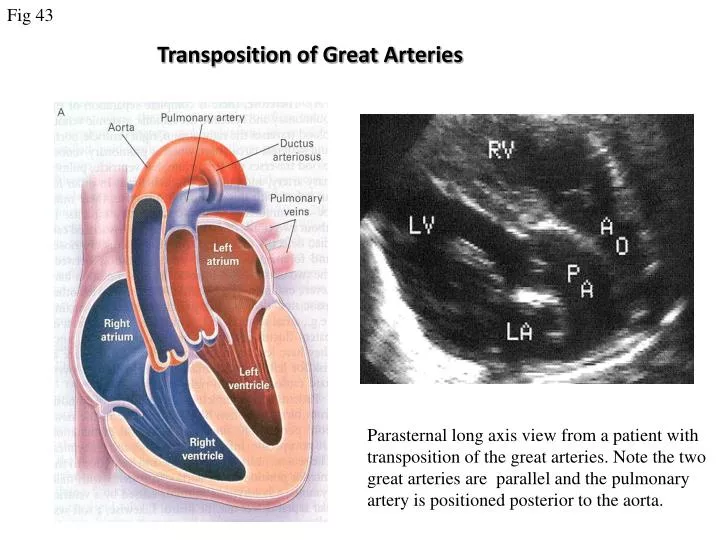
Fig 43. Transposition of Great Arteries. Parasternal long axis view from a patient with transposition of the great arteries. Note the two great arteries are parallel and the pulmonary artery is positioned posterior to the aorta. . Fig 44. RV. LV. Apical four chamber view in a patient with

E N D
Fig 43 Transposition of Great Arteries Parasternal long axis view from a patient with transposition of the great arteries. Note the two great arteries are parallel and the pulmonary artery is positioned posterior to the aorta.
Fig 44 RV LV Apical four chamber view in a patient with transposition of the great arteries after a Mustard repair. Note the intra-atrial baffle (arrow) directing the pulmonary venous flow into the right side atrium. The right atrium and right ventricle are dilated. Mustard Procedure
Fig 45 TGA post Mustard LV RV RA PV Apical four chamber view in a patient with transposition of great arteries after a Mustard repair. Note the intra-atrial baffle (arrow) directing the pulmonary venous flow into the right side atrium. The right atrium and right ventricle are dilated. Colour Doppler mapping demonstrate mild tricuspid regurgitation
Fig 46 TGA post Mustard repair B A mRV mLV C A. Subcostal four chamber view demonstrating pulmonary venous pathway obstruction. B.Colour Doppler flow mapping showing velocity aliasing at the site of stenosis. C. Pulsed wave Doppler trace detecting high flow velocity.
Fig 47 TGA post Mustard repair mLV mRV Apical four chamber view showing severe pulmonary venous pathway obstruction. Note The continuous flow detected at the site of stenosis.
Fig 48 Mustard TR MRV MRV PV atrium Apical four chamber view from a patient with a Mustard repair demonstrating a severely dilated pulmonary venous atrtium which may be the result of severe tricuspid regurgitation and/or impaired right ventricular function. Colour flow map showing the tricuspid regurgitation.