Download
1 / 46
460 likes | 506 Vues
CPAC Rectal Cancer Project. The webinar will begin momentarily. All participant lines will be muted during the presentations. Following the presentations, all participant lines will be unmuted for discussion and question period. CPAC Rectal Cancer Project (2014-2017).

E N D
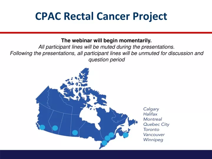
CPAC Rectal Cancer Project The webinar will begin momentarily. All participant lines will be muted during the presentations. Following the presentations, all participant lines will be unmuted for discussion and question period
CPAC Rectal Cancer Project (2014-2017) • Funded by Canadian Partnership Against Cancer (CPAC) • Quality improvement project • 8 centres across Canada • Build multidisciplinary community of practice (COP) to share best practices • Reduce unwarranted variation • Identify gaps in care • Implement strategies to close gaps
This is not QuickSilver! • QuickSilver Study • Phase II study assessing safety and feasibility of using MRI criteria to identify “good prognosis” Stage II and Stage III rectal cancer patients eligible for primary surgery • 17 centres across Canada • Start up meeting June 2013 • Recruitment October 2014 to June 2015
Webinar Outline • Study Overview • Review of process indicators and suggested tools for data capture • Pathology • Radiology • Radiation Oncology • Surgery • Multidisciplinary Cancer Conference • Next Steps and Wrap Up
Objectives - CPAC Rectal Cancer Project To develop a multidisciplinary, pan Canadian Community Of Practice to improve the overall quality of rectal cancer care: • Accelerated diffusion implementation: • Preoperative staging with MRI • Multidisciplinary Cancer Conference (MCC) • High quality radiotherapy and surgical care • High quality pathologic assessment • Select and measure process indicators • Identify gaps in care • Implement strategies to close existing gaps
Year 2 Year 1 Year 3 Apr 2014 - Mar 2015 Apr 2016 – Mar 2017 Apr 2015 – Mar 2016 National Workshop (May 23, 2014) Select and finalize process indicators for each quality initiatives Identify areas for knowledge translation • Planning Phase • (June 1, 2014 to March 31, 2015) • Finalize Project Report Template with final process indicators • Develop tool kit to assist centres capture process indicators • MRI protocol • Synoptic MRI report • Radiation Oncology Pre-Treatment Checklist • Radiation Oncology Treatment Summary • Synoptic OR Report • Pathology Protocol • Pathology CAP Checklist • Develop knowledge translation activities at COP level: • Radiology Training Set • Radiation Oncology Contouring Webinar • Pathology List Serv (EMVI) • Study website and database development • Hire coordinators at each centre (Jan 2015) • Implementation Phase • (April 1, 2015 to Dec 31, 2016) • Audit and Feedback • Measure selected process indicators every 3 months • Generate report (total 7 reports) • Identify gaps and work towards closing gaps • Continue knowledge translation activities on an institutional level FIX THE PROBLEM AGREE WITH CONCEPT FOCUS ON DETAILS
END PRODUCTS • A multidisciplinary model to improve quality of care for rectal cancer • participating centres will be able to take lead to disseminate model in respective provinces • Development of a well coordinated COP • Continue to work together on projects including grant capture and trials
Selection of Process Indicators • Review of pre-workshop survey • Suggested process indicators were rated scale of 1-5 based on clinical importance • National Workshop • Attended by 35 Site and Project Leads; representation from all 8 centres • Discussion of each specific item • Pre-meeting survey results • Best available evidence • Expert opinion • Final vote to include/exclude each specific item • Anonymous; all specialties
Selection of Process Indicators • Results of the final vote reviewed by Investigative Team and Project Leads • Process indicators for which >90% of workshop participants voted to include were kept • Final vote results and final process indicators selected were distributed to meeting participants for final feedback
Suggested Tools and Strategies for Data Capture of Process Indicators • Selected tools and strategies to facilitate data capture based on: • Final process indicators selected • Multidisciplinary discussion at the national workshop • Current best practices at participating centres • Expert and consensus opinion
Suggested Tools and Strategies for Data Capture • Introduce tools selected to facilitate data capture of process indicators • Suggest how tools may be implemented at each centre • Pathology – David Driman • Radiology – Laurent Milot • Radiation Oncology – Charles Cho • Surgery – Lara Williams • MCC – Sender Liberman • Each presentation 10 minutes followed by 5 minutes of questions • Focus questions on suggested tools and implementation • Please direct questions about specific process indicators off line to Site or Project Leads
KEEP IN MIND • All centres receive $2000/site for project launch • All centres encouraged to modify tools or develop new tools to meet their specific institutional needs • All centres to start implementing their plan as soon as possible • All centres receive $60,000/2 years for a research coordinator • All Site Leads will receive a workbook with relevant documents and worksheet to complete (end of November)
CPAC Pathology – Gross Protocol • Specimens grossed as per method of Quirke et al • 3-5 mm slices through fixed, unopened specimen • at least 3 tumour blocks with deepest invasion (CRM where applicable)+ additional tumour block if not already included • at least 2 tumour blocks with closest serosal margin (where applicable) • Macroscopic assessment of quality of the TME • complete, nearly complete, incomplete
CPAC Pathology – Reporting • College of American Pathologists (CAP) checklist - all mandatory elements • College of American Pathologists (CAP) checklist - non-mandatory elements • Assessment of TME quality • Treatment effect following neoadjuvant chemoradiotherapy • Non CAP Elements • Documentation of Quirke method • At least 3 tumour blocks with deepest invasion + 1 tumour block of CRM if not already included • At least 2 tumour blocks with closest serosal surface • Closest distance of CRM in mm (all cases, even when not the closest margin) • Venous (large vessel) invasion – intramural and extramural • Synoptic Format
CPAC Pathology – Practical Issues • Process indicators, protocol and report all selected because of their effect on quality of processes and patient outcomes • A detailed protocol has been developed for each centre to use as a guide • Each centre to determine the best way to implement locally • Report formats can be modified as needed All process indicators will be collected from the pathology report
CPAC Pathology – KT Initiative • Venous invasion selected for KT initiative • VI detection, reporting discussed at national meeting • Encourage use of routine elastin staining • UK guidelines (July 2014) recommend that VI should be detected in at least 30% of resection specimens • Data from reports used to provide audit and feedback to each centre about VI detection rates • Encourage implementation of initiatives to improve detection rates if gaps exist
CPAC MRI Protocol • MRI protocol as per MERCURY • 1.5T or 3.0T magnet with phased array coil (NO endorectal coil) • Includes four fast-spin echo, T2-weighted sequences without fat saturation • High resolution, axial oblique T2 sequences REQUIRED (Sequence 3 and 4) • Optional: • T1 sequences and Diffusion Weighted Imaging • Gadolinium • Bowel preparation • Rectal contrast • Antiperistaltic agents
CPAC Minimum Resolution Requirement • Terminology within protocol can be challenging to interpret • matrix size may be specified as "256 x 128" if there are 128 phase-encoding steps but parallel imaging is used with an acceleration factor of 2 • the spatial resolution would meets requirements however the spatial resolution implied by the scan parameters • Minimum Standard of Resolution Form to be completed by Radiology Site Lead at each centre to ensure that MRI protocol meets this minimum standard of resolution • Each Radiology Site Lead encouraged to work with vendor to verify compliance with protocol • Better resolution (i.e. fewer millimetres per pixel) is acceptable provided SNR is adequately preserved • Generally possible on 3T but harder to accomplish on 1.5T due to time constraints
Benefits of Synoptic MRI Report • Implemented across Ontario in 2009 via Radiology webinar and endorsement by Cancer Care Ontario • A pre and post-implementation audit of 100 MRI reports issued across Ontario showed: • Increase in uptake from 0% to 43% • Increase in completeness from 40% to 79% • Increase in completeness with synoptic compared to narrative format, 95% versus 67% • Increase in distance to to MRF with synoptic compared to narrative format, 84% versus 46%
CPAC MRI KT Initiative • Radiology Training Set developed by Dr Gina Brown • Available on study website in January 2015 • Encourage all participating radiologists to complete 3 MRI reports on-line • Receive Gina Brown’s report to compare your results and access to Gina Brown for questions via an internet chat room • Focus on distance of MRF, EMVI, low rectal cancer, anterior peritoneal reflection
CPAC Radiation Oncology Process Indicators • Process indicators selected because of their effect on quality of processes and patient outcomes • Significiant variation in technique across centres • Peer review of treatment plans is strongly encouraged and may become mandatory • Pre-Treatment Checklist developed to document peer review process • Treatment Summary developed to document treatment details including delivered dose, technique and interruptions in treatment
CPAC Radiation Oncology Process Indicators • Each centre to determine the best way to implement these initiatives locally at their institution • Report format can be modified to best suit needs of each centre • All process indicators will be collected from the Pre-Treatment Radiation Oncology Checklist and the Treatment Summary Form • Both are short, one page forms with check box format • Free text allowed
Radiation Oncology KT Initiative • Recent studies to suggest significant variation in contouring • Planning KT initiative to review current contouring guidelines and assess variation in contouring across centres • Webinar to discuss results, review current guidelines and develop Canadian guideline and develop strategies to minimize variation
Surgery Process Indicators PRE-OPERATIVE ASSESSMENT PROCESS MEASURES SURGICAL PROCESS MEASURES
Benefits of Synoptic OR Report • WebSMR shown to significantly increase the completeness of OR report • Current used in Alberta, Manitoba and Nova Scotia • Uptake low with only 22% of rectal cancer surgeries registered • Likely due to large number of data elements required • Modified version of BC Cancer Agency Rectal Cancer Surgery Checklist • 21 items • Developed to capture all CPAC surgery process indicators
CPAC Surgery Process Indicators and Report • Process indicators and report selected because of their effect on quality of processes and patient outcomes • Synoptic OR report developed as a guide for use at each centre • Each centre determine the best way to implement locally at their institution • Report format can be modified to best suit needs of each centre • All surgery process indicators will be collected OR report • All pre-operative process indicators will be collected from clinical notes in patient chart
Benefits of MCCs • Perceived by health care professionals as improving communication (Devit 2010) • Patients are more likely to receive treatment according to guidelines (McDermid 2009) • Treatment plans will change up to 43% after MCC discussion (Santoso 2004) • May lead to improvements in survival (Prades, 2014)
Results of MCC for Primary Rectal Cancer 31% (13/42) change in initial treatment modality 100% compliance with MCC treatment recommendations Positive CRM rate = 5.5% (2/36)
Synoptic MCC Report • Synoptic MCC report designed to capture selected process indicators • Quality of MCC • Quality of MCC report • Compliance with MCC treatment recommendation • One page in length with check box format • Free text allowed • Format can be modified to suit needs of each centre • Can be completed by MCC Chair and reviewed by treating physician
MCC Suggested KT Initiative • In addition to study outcomes: • Quality of MCC • Quality of the MCC report • Compliance with MCC treatment recommendation • Complete TME and positive CRM rate • Encourage each centre to assess the change in the initial treatment modality over a six month time period during the study
SUMMARY AND NEXT STEPS • Site Leads at each centre to launch project locally ($2000 per site) • Review process indicators and best local strategies to ensure capture of the indicators • Surgical Site Leads to ensure REB and DSA complete • Transfer of $30K (Jan 2015 and Jan 2016) • Hire coordinator (consider using pre-existing coordinators)
SUMMARY AND NEXT STEPS • Start to implement initiatives as soon as possible • Patient recruitment/data collection begins April 1, 2015 (first report July 2015) • Regular teleconference last Tuesday every other month starting January 27th • Newsletter with meeting minutes • Website for on-site data collection • Webinar for research coordinators in March 2015 to review definitions/terms for data collection
THANK YOU! • REB/DSA (Surgical Site Lead) • Hire Coordinator for January 2015 (Surgical Site Lead) • Project Launch as soon as possible (All Site Leads) • Complete Worksheet (All Site Leads) • Next teleconference: (All Site and Project Leads) Tuesday January 27th from 4:30-5:30 pm EST