Download
1 / 45
450 likes | 608 Vues
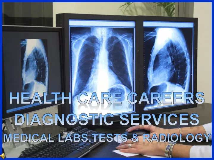
HEALTH CARE CAREERS. DIAGNOSTIC SERVICES. MEDICAL LABS,TESTs & Radiology. The medical laboratory… phlebotomy technician. Many diagnostic tests require a blood sample. These may be collected by a phlebotomy technician that has been trained on-the-job or in a formal training program.

E N D
HEALTH CARE CAREERS DIAGNOSTIC SERVICES MEDICAL LABS,TESTs & Radiology
The medical laboratory… phlebotomy technician Many diagnostic tests require a blood sample. These may be collected by a phlebotomy technician that has been trained on-the-job or in a formal training program. They most commonly obtain a blood sample through a procedure called venipuncture (VEEN ah punk chur), the puncturing of a vein with a needle designed for blood collection.
The medical laboratory… phlebotomy technician The phlebotomist must interact with the client, reassuring them, maintaining confidentiality, and answering their questions. They must explain the procedure, correctly obtain and label the samples, and transport specimens to the designated location.
The medical laboratory… medical lab assistant (MLA) The medical lab assistant can perform the tasks of the phlebotomist, plus more complex tests such as urine or throat cultures, giving injections, cholesterol and blood glucose tests, electrocardiograms, preparing bacteriological smears, and taking vital signs…all used to help the physician make a diagnosis.
The medical laboratory… medical lab technician (MLT) The medical lab technician usually holds a two-year degree and has passed a national certification test. They can collect the samples, but more often only do the analysis on the samples. They prepare blood, tissue, cell, or other body fluid specimens and operate automated analyzers. They must keep accurate records.
The medical laboratory… clinical laboratory scientist (CLS) The medical technologist or clinical laboratory scientist has a 4 year degree in medical technology or life sciences. They can perform all types of tests, looking for bacteria, parasites, and other microorganisms, analyzing the chemical content of fluids, testing for drug levels in the blood, preparing specimens, counting cells, or looking for abnormal cells.
The medical laboratory… inoculating an agar plate ‘Inoculating an agar plate’ means to grow microorganisms into a culture on a petri dish using agar (AH gur). Agar supplies the nutrients that enable microorganisms to grow. Once the microorganism is isolated, it is tested for sensitivity to specific antibodies. This allows us to identify it.
The medical laboratory… preparing a bacteriological smear A bacteriological smear is used to prepare a specimen of throat, wound, urine, sputum, and skin tissues. The specimen is collected on a swab, swiped onto a glass slide, air-dried and heat-fixed. It is now ready for a staining procedure that can identify microorganisms that may be present.
The medical laboratory… staining bacteriological smears Adding a colored stain to a biological smear helps identify the microorganisms that may be present. ‘Grams stain’ is the most frequently used stain. Gram-positive Smears that retain the violet color of the gram’s stain (gram-positive) are caused by bacteria with thick walls. Smears that turn red when exposed to the gram’s stain (gram-negative) are caused by bacteria with thin walls. This is an initial step in identifying the type of an infection.
The medical laboratory… measuring specific gravity of urine A urinometer is used to measure the specific gravity of urine. Solids in the urine, such as cells or crystals, increase specific gravity. Specific gravity is higher first thing in the morning than after a day of drinking fluids. A urinometer used in distilled water reads 1.000; normal specific gravity for urine is 1.010-1.025. Abnormally high specific gravity may indicate urinary tract infections, dehydration, adrenal insufficiency, liver disease, congestive heart disease, or diabetes.
The medical laboratory… testing urine with reagent strips Plastic strips that have been treated with various reagents (rē-ā-jənts) are used as dipsticks in urine. The color on the strip changes to indicate the presence or specific amounts of certain substances. A viewer is used to help accurately read the test strips.
The medical laboratory… testing urine with reagent strips Reagent strips test urine for: bilirubin(liver disease) blood (infections or kidney bleeding) glucose(diabetes) ketones (prolonged fasting) leukocytes(white blood cells indicating infections) nitrites(bacterial infections) pH(metabolic, respiratory, or urinary tract problems) protein(renal disease,heavy exercise, high fever) human chorionic gonadotropin – HCG(pregnancy)
Medical testing… the electrocardiography technician An electrocardiography technician uses a machine to record electrical activity of the heart. This record, referred to as an ECG or EKG, is called a ‘tracing’ and helps the physician diagnose and evaluate cardiovascular disease. Accuracy may be compromised by electrical interference or defective ‘leads’. The technician is trained on-the-job or in a certification program.
Medical testing… the electrocardiography technician Exercise electrocardiography, commonly called a stress test, measures the heart’s activity while the client walks on a treadmill, pedals an exercise bike, or walks up a flight of stairs. The technician must monitor blood pressure, heart rate, skin temperature, oxygen levels, and physical appearance during this test… and be ready to handle an emergency.
Medical testing… the cardiovascular technologist With an additional 2-4 years of training, the electrocardiography technician can become a cardiovascular technologist. They can assist with more invasive tests such as angioplasty (expands a blocked artery), cardiac catherization (a dye is injected through a tube from the groin into the heart), heart surgery, or insertion of pacemakers.
Medical testing… the cardiovascular technologist The cardiovascular technologist may specialize in performing ultrasounds of the heart, known as echocardiograms. during this test… and be ready to handle an emergency.
Medical testing… the electroencephalography (EEG) and electroneurodiagnostic (END) technologists These technologists are trained on-the-job or in 1-2 year programs. Each places ‘leads’ or ‘electrodes’ on the client, and records a tracing of electrical impulses. The EEG technologist records the electrical activity of the brain. The END technologist records the electrical activity of the nervous system.
Radiology… We’ve already talked about the physician who specializes in radiology. This doctor relies upon images to diagnose or treat diseases, and is assisted by numerous other technologists and therapists. They may specialize in diagnostic radiology (diagnoses disease or injury) or radiation oncology (uses radiation in the treatment of cancer).
Diagnostic radiology… The radiologic technologist, nuclear medicine technologist, or radiation therapy technologist usually has 1-4 years of training leading to a degree and/or certification. Registered technologists must always continue their education to stay certified.
Diagnostic radiology… The radiologic technologist assists clients before, during, and after procedures, operates equipment, maintains aseptic and sterile techniques and standard precautions, uses proper shielding from the radiation, positions clients comfortably during procedures, processes and records film or images for the radiologist.
Diagnostic radiology… Radiology workers rely upon various imaging methods or modalities. Invasive images are taken from inside the body, requiring the insertion of catheters, drugs, or contrast media such as organic iodine or barium. Noninvasive images are taken from outside the body and use no contrast mediums (injected dyes).
Diagnostic modalities… radiography The majority of radiologic technologists are employed as radiographers. Short electromagnetic waves called X-rays are used to generate radiographs… two-dimensional images of light and dark shadows on firm, computer disk, or videotape.
Diagnostic modalities… fluoroscopy A fluoroscopy is achieved by projecting an Xray image onto a fluorescent screen for viewing. The use of an invasive contrast medium makes the image easier to see. A catheter can be advanced through the artery and into the heart, and the contrast medium is injected through the catheter. This method is used for fluoroscopy images called angiograms (images of arteries and veins) and arteriograms (images of arteries). Barium may be swallowed or injected into the rectum for gastro-intestinal exams.
Diagnostic modalities…computed tomography A computed tomography technologist uses an Xray machine called a computed axial tomography scanner… or CAT scan. The images view the body in slices, much like the slices in a loaf of bread.
Diagnostic modalities… sonography Sonographers use a transducer to pass high-frequency sound waves through the body. Echos bounce back from internal organs or tissues, and are converted to images. This is commonly called an ultrasound procedure.
Diagnostic modalities… magnetic resonance imaging Magnetic resonance imaging (MRI) uses a large electromagnet to subject the body to a strong magnetic field that aligns the hydrogen atoms in the body and produces an image that is clearer than radiography or CAT scans. It is important that clients do not have metal objects, such as metal plates, in their body.
A nuclear medicine technologist injects or attaches radioactive isotopes to the blood. A multigated acquisition or MUGA scan can image heart function. Positron emission tomography or PET scan can image blood flow or brain activity. Radioactive iodine uptake images thyroid function. Diagnostic modalities… nuclear medicine PET PET MUGA
Diagnostic modalities… mammography Mammography technologists use special machines to produce diagnostic images of breast tissue, helping in the early detection and treatment of breast cancer. This improves survival rates.
Picture archival and communications systems… PACS When diagnostic images are digitalized, they can be viewed, stored, transmitted, archived, and retrieved on computers. This allows the entire health care team access, and is an important component of information and insurance/medical cost management.
Radiation oncology… The radiation oncologist is assisted by the radiation therapist. The therapist administers radiation doses that target cancer cells in the client’s body. This stops or slows their growth.
Radiation oncology… Several types of radiation therapies are common. External beam therapy involves the daily delivery of radiation via cobalt, over a period of 4-8 weeks. Brachytherapy (break ee therapy) treats tumors inside the body. Radioactive seeds are inserted directly into or next to the tumor to shrink it. Linear accelerator (LINAC) uses microwave technology to produce more powerful Xrays to destroy cancer cells.
Protection from radiation… Exposure to radiation causes cell damage, so the technologists must protect themselves. Radiation can bounce or scatter, so a distance of 6 feet from the radiation source is recommended. Protective barriers often contain lead, which radiation cannot penetrate. They can be used to target the radiation rays, or cover organs, or line walls and gloves. Radiation badge detects level of exposure
THE END HEALTH CARE CAREERS DIAGNOSTIC SERVICES MEDICAL LABS,TESTs & Radiology
What do you see in this x-ray? Ovarian cyst – a cyst shows up as solid black on ultrasound
What do you see in this x-ray? Two broken bones
What do you see in this ultrasound? The yolk sacs and umbilical cords in an early pregnancy of twins
One broken bone in the finger What do you see in this x-ray?
What do you see in this digital image? The functioning of a heart
Can you identify the following conditions in these digital images? early pregnancy, single broken bone, 2 broken bones, ovarian cyst, heart function