Download
1 / 32
330 likes | 377 Vues
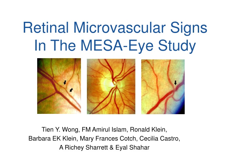
Retinal Microvascular Signs In The MESA-Eye Study. Tien Y. Wong, FM Amirul Islam, Ronald Klein, Barbara EK Klein, Mary Frances Cotch, Cecilia Castro, A Richey Sharrett & Eyal Shahar. Historical Perspective Retinal microvascular signs were described in in patients with severe hypertension.

E N D
Retinal Microvascular Signs In The MESA-Eye Study Tien Y. Wong, FM Amirul Islam, Ronald Klein, Barbara EK Klein, Mary Frances Cotch, Cecilia Castro, A Richey Sharrett & Eyal Shahar
Historical PerspectiveRetinal microvascular signs were described in in patients with severe hypertension • Hypertensive retinopathy classification paper by Keith, Wagener and Barker in 1941 • Keith NM, Wagener HP, Barker NW. Am J Med Sci 1939; 197:332-43. • Marcus Gunn described these signs in a series of patients with kidney disease in 1898 • Gunn RM. Trans Ophthalmol Soc U K 1892; 12:124-5.
Retinal Signs & Incident StrokeThe ARIC study Wong TY, Klein R, Couper DJ, et al. Lancet 2001: 358: 1134-40
Retinal Signs & Incident CHFThe ARIC study Wong TY, Rosamond WR, Chang P, et al. JAMA 2005; 293:63-69
Retinal AVR & Incident DMAVR reflects relative caliber of retinal arterioles/venules ARIC Study 3-yr DM incidence (Wong et al JAMA 2002) Beaver Dam Study 10-yr DM incidence (Wong et al Arch Intern Med 2005)
Objectives • Describe prevalence and distribution of retinal microvascular signs in the MESA-Eye Study • Examine associations of retinal microvascular signs with both conventional and novel cardiovascular risk factors, including biomarkers of inflammation, hemostasis & endothelial dysfunction • Examine possible ethnic/racial variations
Assessment of Retinal Microvascular Signs • Non-mydriatic digital retinal photographs of both eyes • Two photographs per eye (optic disc and macula) • Evaluated at University of Wisconsin, Madison • Focal retinal signs (Two eyes) • Focal arteriolar narrowing • Arterio-venous (AV) nicking • Retinopathy (microaneurysm, hemorrhages, cotton wool spots) • Retinal vascular caliber (One eye) • Retinal arteriolar caliber (CRAE) • Retinal venular caliber (CRVE)
Microaneurysm Focal narrow AV nicking
Cotton wool spots AV nicking
Study Population 6,814 MESA Baseline Exam 6,239 MESA 2nd Exam • 6,147 Retinal photographs • 6,106 AV nicking • 6,070 Focal narrowing • 5,282 Retinopathy (no DM) • 5,979 Retinal vascular caliber
Retinal Vascular Caliber Arteriolar Caliber Venular Caliber
Retinal Vascular Caliber Arteriolar Venular DBP SBP
Retinal Vascular Caliber Arteriolar Venular Fasting glucose BMI
Markers of Inflammation Arteriolar Venular IL-6 Plasma Fibrinogen CRP
No Associations • Past medical history of inflammatory disease • Asthma, arthritis, and hepatitis, • Medication use • Lipid lowering agents, diuretics, oral contraceptives, HRT, oral steroids, aspirin and NSAIDs, • Hematology/Biochemistry • TNF- • Soluble E-selectin, vWF, factor VIII • Plasmin-antiplasmin complex, D-dimer, total homocysteine, soluble tissue factor, thrombin activatable fibrinolysis inhibitor, tissue factor pathway inhibitor, soluble thrombomodulin, anti-human heat shock protein 60, remnant-like particle cholesterol • Interleukin-2 soluble receptor chain, Matrix metalloproteinase 3, matrix metalloproteinase 9, • IgG to Chlamydia pneumoniae, Herpes simplex virus, Cytomegalovirus, Helicobacter pylori and Hepatitis A
Ethnic/Racial Variations Focal Retinal Signs Model 1: age, gender and center Model 2: Model 1 variables plus for AV nicking: SBP, hypertension, DM, asthma, smoking, glucose, BMI, waist hip ratio, HDL, triglycerides, plasmin-antiplasmin complex, hsCRP, interleukin-6 and education; for focal narrowing: SBP, hypertension, DM, gum disease, WHR, TG, and education and for retinopathy: SBP and hypertension, smoking, glucose, BMI and education.
Ethnic/Racial Variations Retinal Vascular Caliber Mean difference (95% confidence interval) of arteriolar and venular diameter, comparing Blacks, Hispanics or Chinese with Whites, adjusted for age, gender and center † For arteriolar caliber, additional adjustment for systolic blood pressure, hypertension, diabetes, smoking, alcohol consumption, BMI, plasma fibrinogen, total homocysteine and education; for venular caliber, additional adjustment for systolic blood pressure, diabetes, smoking, BMI, waist hip ratio, glucose, triglycerides, LDL and HDL cholesterol, hsCRP, plasma fibrinogen, interleukin-6 and education.
Possible Explanations • Racial/ethnic differences in the distribution of CVD risk factors • Blacks and Hispanics were more likely to have DM, obesity, dyslipidemia and systemic inflammation These factors are associated with larger venular caliber But they did not explain the observed racial differences in adjusted models • Racial/ethnic differences in the susceptibility and manifestations of retinal microvasculature to common CVD risk factors? • Differences in refractive errors? • Myopic refraction associated with smaller retinal vascular caliber • Whites and Chinese have a higher prevalence of myopia…
Pathophysiology Diabetes Retinal Venular Caliber Retinopathy Retinal Microvascular Signs Metabolic syndrome Inflammation Endothelial dysfunction? Retinal Arteriolar Caliber AV nicking Hypertension
Conclusion • Different associations of retinal microvascular signs with standard and novel CVD risk factors • AV nicking, focal narrowing, retinopathy associated with hypertension • AV nicking associated with inflammation • Retinal arteriolar caliber is associated with hypertension • Retinal venular caliber is associated with diabetes, dyslipidemia, obesity and diabetes • Findings may provide insights into the different systemic processes associated with retinal microvascular signs and why they predict CVD events
Future Analyses • Associations with subclinical CVD • CAC and carotid IMT • LV mass and function • Major and minor ECG abnormalities • Endothelial function (?) • Associations with other measures in MESA • Lipoprotein subclass/particle size/oxidized LDL • Renal function • Cognitive function, depression • Air pollution • Other Eye endpoints • Age-related macular degeneration • Diabetic Retinopathy • Vein occlusions, epiretinal membranes and other retinal diseases • Refraction • Visual impairment • Associations with incident CVD events…