Download
1 / 21
210 likes | 436 Vues
Airway and Ventilatory Management. Presented by: Barry Nicholson, M.D. Scenario –.

E N D
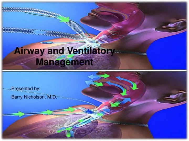
Airway and Ventilatory Management Presented by: Barry Nicholson, M.D.
Scenario – A 22 year-old male is an unrestrained passenger in a motor vehicle that collides head-on into retaining wall. He has a strong odor of alcohol on his breath. At the time of the collision, he hits the windshield and sustains a scalp laceration. At the injury scene, he is combative, and his GCS score is 11. His blood pressure is 120/70 mm Hg, his heart ate is 100 beats/min, and his respirations are 38 breaths/min and O2 saturation is 90%. A semi rigid cervical collar is applied, and he is immobilized on a long backboard. He is receiving oxygen via a high-flow oxygen mask. Shortly after his arrival in the ER he begins to vomit.
Objectives: • Identify clinical situations in which airway compromise is likely to occur. • 2. Recognize the signs and symptoms of acute airway obstruction.
3. Techniques for establishing and maintaining a patient’s airway. 4. Techniques for confirming the adequacy of ventilation and oxygenation.
Supplemental oxygen must be administered to all trauma patients. • Airway compromise and signs of airway obstruction: • Maxillofacial trauma-Hemorrhage, increased secretions, dislodged teeth, fractured mandible. • Patients who refuse to lie down, may be experiencing difficulty in maintaining their airway. Agitation, cyanosis, retractions, noisy breathing suggests hypoxemia. • Neck trauma-displacement and obstruction of the airway. Surgical airway. • Laryngeal trauma-triad. • 1.Hoarsness 2. Subcutaneous emphysema 3. Palpable fracture.
Ventilation • chest trauma • CNS compromise • pulmonary dysfunction
AirwayManagement Look: rise and fall of chest, Tachypnea, accessory muscle use, pulse oximeter Listen: decreased or absent breath sounds, noisy breathing Feel: rib fractures, subcutaneous emphysema -chin lift -jaw thrust
Definitive Airway – • Apnea • Inability to maintain a patent airway by other means. • Protect the lower airway from aspiration • Impending or potential compromise-seizure, burns, facial fractures, retropharyngeal hematoma • Glascow Coma scale <8 • Inadequate oxygen saturation with facemask
Endotracheal Intubation VIDEO Click for Video demonstration If Hyperlink fails: go to http://www.youtube.com/watch?v=tKz2zadEX_0
Steps 1 & 2– Step 1: Ensure that adequate ventilation and oxygenation are in progress and that suctioning equipment is immediately available in the event the patient vomits. Step 2: Inflate the cuff of the endotracheal tube to ascertain that he balloon does not leak and then deflate the cuff.
Steps 3, 4 & 5 Step 3: Connect the laryngoscope blade to the handle and check the bulb for brightness Step 4: Assess the patient’s airway for ease of intubation (LEMON mnemonic) Step 5: Direct an assistant to manually immobilize the head and neck. The patient’s neck must not be hyper-extended or hyper-flexed during the procedure
Steps 6 thru 9 Step 6: Hold the laryngoscope in the left hand. Step 7: Insert the laryngoscope into the right side of the patient’s mouth, displacing the tongue to the left. Step 8: Visually indentify the epiglottis and then the vocal cords. Step 9: Gently insert the endotracheal tube into the trachea without applying pressure on the teethe or oral tissues.
Steps 10, 11 & 12 Step 10: Inflate the cuff with enough air to provide an adequate seal. Do NOT over-inflate the cuff. Step 11: Check the placement of the endotracheal tube by bag-mask-to-tube ventilation. Step 12: Visually observe chest excursions with ventilation.
Steps 13 & 14 Step 13: Auscultate the chest and abdomen with a stethoscope to ascertain the tube position. Step 14: Secure the tube. If the patient is moved, the tube placement should be reassessed.
Step 15 Step 15: If endotracheal intubation is not accomplished within seconds or in the same time required to hold your breath before exhaling, discontinue attempts, apply ventilation with a bag-mask device and then try using the gum elastic bougie.
Steps 16 & 17 Step 16: Placement of the tube must be checked carefully. A chest x-ray exam is helpful to assess the position of the tube, but it cannot exclude esophageal intubation Step 17: Attach a CO2 detector to the endotracheal tube between the adapter and the ventilating device to confirm the position of the endotracheal tube in the airway.
Step 18 Attach a pulse oximeter to one of the patient’s fingers (intact peripheral perfusion must exist) to measure and monitor the patient’s oxygen saturation levels and provide an immediate assessment of therapeutic interventions.
Reviewing the Scenario – A 22 year-old male is an unrestrained passenger in a motor vehicle that collides head-on into a retaining wall. He has a strong odor of alcohol on his breath. At the time of the collision, he hits the windshield and sustains a scalp laceration. At the injury scene, he is combative, and his GCS score is 11. His blood pressure is 120/70 mm Hg, his heart rate is 100 beats/min, and his respirations are 38 breaths/min and O2 saturation is 90%. A semi rigid cervical collar is applied, and he is immobilized on a long backboard. He is receiving oxygen via a high-flow oxygen mask. Shortly after his arrival in the ER he begins to vomit.
Airway and Ventilatory Management Presented by: Barry Nicholson, M.D.