Download
1 / 69
690 likes | 693 Vues
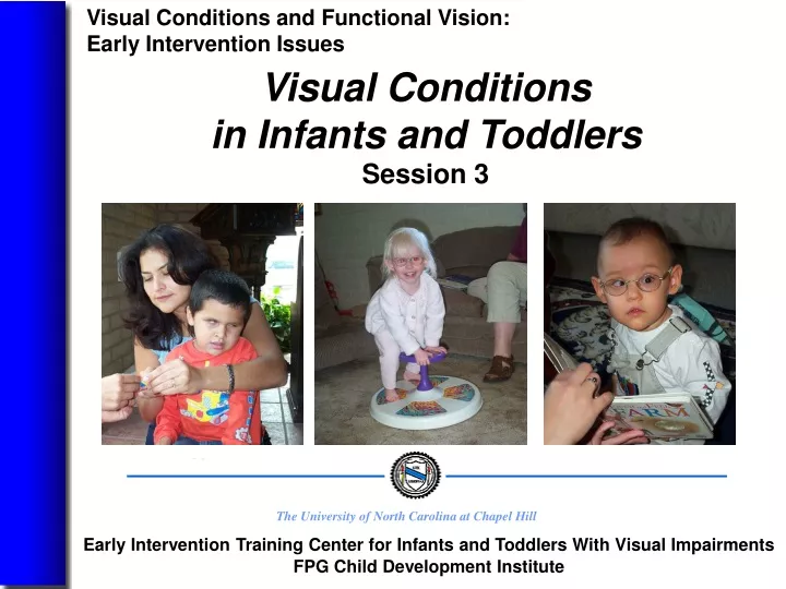
This session explores the most prevalent visual conditions in infants and toddlers with visual impairments, including cortical visual impairment, retinopathy of prematurity, optic nerve hypoplasia, and structural abnormalities. Participants will learn about causes, characteristics, and implications of each condition for early development and intervention.

E N D
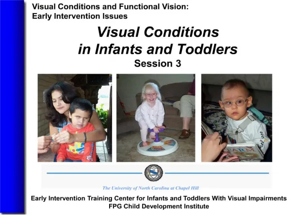
Visual Conditions and Functional Vision: Early Intervention Issues Visual Conditions in Infants and ToddlersSession 3 The University of North Carolina at Chapel Hill Early Intervention Training Center for Infants and Toddlers With Visual Impairments FPG Child Development Institute
After completing this session, participants will 1. identify the most prevalent visual conditions found in young children with severe visual impairments in the United States and Canada and how they differ from those found in adults. Objectives Early Intervention Training Center for Infants and Toddlers with Visual Impairments FPG Child Development Institute University of North Carolina at Chapel Hill June 4, 2004 Visual Conditions 3A
After completing this session, participants will identify the three most prevalent visual conditions—cortical visual impairment (CVI), retinopathy of prematurity (ROP), and optic nerve hypoplasia (ONH)—in young children with visual impairments. Describe causes and characteristics of each condition as well as the implications for early development and intervention. Objectives Early Intervention Training Center for Infants and Toddlers with Visual Impairments FPG Child Development Institute University of North Carolina at Chapel Hill June 4, 2004 Visual Conditions 3B
After completing this session, participants will discuss the causes, characteristics, and implications of the following visual conditions: structural abnormalities—anophthalmia, microphthalmia, coloboma, albinism, retinal disorders such as retinoblastoma and Leber’s Congenital Amaurosis, congenital cataracts, and delayed visual maturation. Objectives Early Intervention Training Center for Infants and Toddlers with Visual Impairments FPG Child Development Institute University of North Carolina at Chapel Hill June 4, 2004 Visual Conditions 3C
After completing this session, participants will 4. describe the characteristics and implications of the following conditions that may occur as primary or secondary diagnoses—strabismus, amblyopia, glaucoma, nystagmus, and refractive errors. Objectives Early Intervention Training Center for Infants and Toddlers with Visual Impairments FPG Child Development Institute University of North Carolina at Chapel Hill June 4, 2004 Visual Conditions 3D
The prevalence of severe visual impairments in developing countries is about 1 in 1,000, as compared to about 1 in 10,000 in wealthy countries. The most prevalent visual conditions in adults with severe visual impairments are diabetic retinopathy, macular degeneration, cataracts, and glaucoma. Hatton and colleagues (2001) reported that the most prevalent visual conditions in young children in their sample were CVI, ROP, ONH, albinism, and structural abnormalities such as anophthalmia, microphthalmia, and coloboma. Prevalence of Visual Impairments Early Intervention Training Center for Infants and Toddlers with Visual Impairments FPG Child Development Institute University of North Carolina at Chapel Hill June 4, 2004 Visual Conditions 3E
DiagnosisReferral CVI 7.9 months 10.9 months ROP 2.4 months 11.5 months ONH 4.3 months 8.1 months Structural 2 weeks 9.5 months Albinism 3.4 months 11.7 months Other 5.2 Months 11.3 months Critical Events in Visual Conditions:Age of Diagnosis Hatton et al., 2001 Early Intervention Training Center for Infants and Toddlers with Visual Impairments FPG Child Development Institute University of North Carolina at Chapel Hill June 4, 2004 Visual Conditions 3F
Structural abnormalities may be diagnosed very early because they may be apparent soon after birth. Lag time between diagnosis and referral suggests that closer collaboration with eye care specialists and other early intervention programs is needed. Earlier referral could lead to more immediate supports for families and facilitation of optimal development of infants with VI. Hatton et al., 2001 Diagnosis and Referral Early Intervention Training Center for Infants and Toddlers with Visual Impairments FPG Child Development Institute University of North Carolina at Chapel Hill June 4, 2004 Visual Conditions 3G
Hatton et al. (2001) reported that the most prevalent visual conditions in a sample of 406 infants and toddlers with severe VI were cortical visual impairment (CVI), retinopathy of prematurity (ROP), optic nerve hypoplasia (ONH), structural abnormalities, and albinism. This was consistent with studies reported by Ferrell (1998), Hatton (1991); Hatton et al. (1997), and Steinkuller et al. (1999). Most Prevalent Conditions in Young Children With Severe VI Early Intervention Training Center for Infants and Toddlers with Visual Impairments FPG Child Development Institute University of North Carolina at Chapel Hill June 4, 2004 Visual Conditions 3H
It is difficult to determine whether infants and toddlers meet criteria for legal blindness. Approximately 63% of children with structural abnormalities and 42% of children with albinism were designated legally blind in the Hatton et al. study (2001). Children with diagnoses of legal blindness may have access to more resources, for example, quota funds for developmental resources from the American Printing House for the Blind. Amount of Vision in Young Children With Severe VI Early Intervention Training Center for Infants and Toddlers with Visual Impairments FPG Child Development Institute University of North Carolina at Chapel Hill June 4, 2004 Visual Conditions 3I
Children with albinism are more likely to have a single disability of visual impairment when enrolled in specialized programs (Hatton et al., 2001). Children with CVI are most likely to have additional disabilities at time of enrollment in specialized programs for children with VI (Hatton et al., 2001). Children with multiple disabilities and their families may require supports and services that are specific to their unique needs based on each child’s combination of disabilities. Multiple Disabilities and VI Early Intervention Training Center for Infants and Toddlers with Visual Impairments FPG Child Development Institute University of North Carolina at Chapel Hill June 4, 2004 Visual Conditions 3J
Children with CVI and ROP are more likely to have co-occurring health conditions. Infants and toddlers with CVI and ROP who depend on technology may have unique medical needs that affect early intervention. Some sensory stimulation activities may trigger seizures. Children with respiratory problems may be sick more often and more likely to catch contagious illnesses. Health Conditions and VI Early Intervention Training Center for Infants and Toddlers with Visual Impairments FPG Child Development Institute University of North Carolina at Chapel Hill June 4, 2004 Visual Conditions 3K
Ferrell (1998) and Hatton et al. (2001) found CVI to be the most prevalent visual condition in young children with severe VI. CVI results from injury to the brain or visual pathways in the brain rather than disorders or abnormal structures of the eye. CVI varies in severity from child to child and from environment to environment, and children with CVI may experience improvement in visual function. Cortical Visual Impairment (CVI) Early Intervention Training Center for Infants and Toddlers with Visual Impairments FPG Child Development Institute University of North Carolina at Chapel Hill June 4, 2004 Visual Conditions 3L
Oxygen deprivation (hypoxia, ischemia) Prematurity Periventricular leukomalacia Trauma Meningitis Causes of CVI Early Intervention Training Center for Infants and Toddlers with Visual Impairments FPG Child Development Institute University of North Carolina at Chapel Hill June 4, 2004 Visual Conditions 3M
CVI can be divided into two groups: cortical and subcortical injuries. Cortical Subcortical • exotropia • esotropia • horizontal conjugate • tonic downgaze gaze deviation • ONH and other optic nerve abnormalities Children in both groups have roving eye movements associated with severe visual impairment and similar rates of nystagmus. Brodsky et al., 2003 Visual Behaviors and CVI Early Intervention Training Center for Infants and Toddlers with Visual Impairments FPG Child Development Institute University of North Carolina at Chapel Hill June 4, 2004 Visual Conditions 3N
Children with CVI typically have neurological abnormalities in addition to other ocular disorders, fluctuating vision based on fatigue and levels of sensory input, limited or no eye contact, vision that generally improves over time but does not extend to typical levels of vision, and rates of improvement that are determined by the age at which CVI occurred and the area of the brain that is injured. Carden & Good, 2003 Visual Behaviors and CVI Early Intervention Training Center for Infants and Toddlers with Visual Impairments FPG Child Development Institute University of North Carolina at Chapel Hill June 4, 2004 Visual Conditions 3O
The following characteristics have been documented in children with CVI: additional neurological abnormalities, fluctuations in vision, preferences for colored objects, light gazing, and turning head and eyes away from objects while reaching for them. Good et al., 1994 Jan et al., 1987 Visual Behaviors and CVI Early Intervention Training Center for Infants and Toddlers with Visual Impairments FPG Child Development Institute University of North Carolina at Chapel Hill June 4, 2004 Visual Conditions 3P
The following characteristics have been documented in children with CVI: using touch rather than vision to identify objects, preference for familiar environments, and photophobia in about a third of children with CVI. Good et al., 1994 Jan et al., 1987 Visual Behaviors and CVI Early Intervention Training Center for Infants and Toddlers with Visual Impairments FPG Child Development Institute University of North Carolina at Chapel Hill June 4, 2004 Visual Conditions 3Q
In a sample of 406 children, 86 had CVI. Approximately half of children with CVI were considered legally blind. 79% appeared to have developmental delays or multiple impairments. 57% had seizures. 24% had eating disorders. 21% were dependent on technology (e.g., tracheotomies or GI tubes). 17% had respiratory problems. Hatton et al., 2001 Characteristics of Children With CVI Early Intervention Training Center for Infants and Toddlers with Visual Impairments FPG Child Development Institute University of North Carolina at Chapel Hill June 4, 2004 Visual Conditions 3R
The prevalence of ROP has increased since the 1980s because improved technology has allowed smaller and younger infants to survive. ROP is responsible for 500 to 550 new cases of blindness in the U.S. each year (Siatkowski & Flynn, 1998). Medical technology constantly evolves, making it challenging to stay abreast of the latest trends in treatment. Retinopathy of Prematurity (ROP) Early Intervention Training Center for Infants and Toddlers with Visual Impairments FPG Child Development Institute University of North Carolina at Chapel Hill June 4, 2004 Visual Conditions 3S
The premature infant’s eye with ROP has a layer of blood vessels in the retina that have grown excessively, forming a ridge of scar tissue over the retina and affecting visual function. Premature Eye With ROP IRIS Medical. (1991). Understanding retinopathy of prematurity (p. 5) [Brochure]. Mountain View, CA: IRIS Medical Instruments, Inc. Used with permission. Early Intervention Training Center for Infants and Toddlers with Visual Impairments FPG Child Development Institute University of North Carolina at Chapel Hill June 4, 2004 Visual Conditions 3T
ROP is classified by the zones of the eye that it affects. Zone 1 encompasses the optic nerve and the macula. Zone 2 includes the optic nerve, the macula, and a larger portion of the eye. Zone 3 encompasses all regions of the eye, including the ora serrata. Classification of ROP Scheme of retina IRIS Medical. (1991). Understanding retinopathy of prematurity (p.6) [Brochure]. Mountain View, CA: IRIS Medical Instruments, Inc. Used with permission. Early Intervention Training Center for Infants and Toddlers with Visual Impairments FPG Child Development Institute University of North Carolina at Chapel Hill June 4, 2004 Visual Conditions 3U
The location of the disease is denoted by zones. Zone I: The inner zone extends from the optic disc to twice the disc-macular distance, or 30 degrees in all directions from the optic disc. Zone II: The middle zone extends from the outer border of Zone I to the ora on the nasal side and to approximately the equator on the temporal side. Zone III: The outer zone extends from the outer edge of Zone II in a crescentic fashion to the ora serrata. Flynn, 1991, p. 64 Classification of ROP Early Intervention Training Center for Infants and Toddlers with Visual Impairments FPG Child Development Institute University of North Carolina at Chapel Hill June 4, 2004 Visual Conditions 3V
Stage 1: A thin, relatively flat, white demarcation line separates the avascular retina anteriorly, from the vascularized retina posteriorly. Vessels that lead up to the demarcation line are abnormally branched and/or arcaded. Ober et al., 2003, p. 602 Stages of ROP Early Intervention Training Center for Infants and Toddlers with Visual Impairments FPG Child Development Institute University of North Carolina at Chapel Hill June 4, 2004 Visual Conditions 3W
Stage 2: The demarcation line has visible volume and extends off the retinal surface as a white or pink ridge. Retinal vessels may appear stretched locally, and vault off the surface of the retina to reach the peak of the ridge. Tufts of neo-vascular tissue may be present posterior to, but not attached to, the ridge. Ober et al., 2003, p. 602 Stages of ROP Early Intervention Training Center for Infants and Toddlers with Visual Impairments FPG Child Development Institute University of North Carolina at Chapel Hill June 4, 2004 Visual Conditions 3X
Stage 3: Extraretinal fibrovascular (neovascular) proliferative tissue emanates from the surface of the ridge, extending posteriorly along the retinal surface, or anteriorly toward the vitreous cavity, giving the ridge a ragged appearance. Ober et al., 2003, p. 602 Stages of ROP Early Intervention Training Center for Infants and Toddlers with Visual Impairments FPG Child Development Institute University of North Carolina at Chapel Hill June 4, 2004 Visual Conditions 3Y
Stage 4: Subtotal retinal detachment. Traction type retinal detachment results from the development of proliferating tissue in the vitreous gel or on retinal surfaces, subdivided into two types. Ober et al., 2003, p. 602 Stages of ROP Early Intervention Training Center for Infants and Toddlers with Visual Impairments FPG Child Development Institute University of North Carolina at Chapel Hill June 4, 2004 Visual Conditions 3Z
4A. Subtotal retinal detachment not involving the fovea that generally carries a relatively good prognosis because the macula and fovea are not affected. 4B. Subtotal retinal detachment involving the fovea and macula that results in poor vision. Flynn, 1991 Stages of ROP Early Intervention Training Center for Infants and Toddlers with Visual Impairments FPG Child Development Institute University of North Carolina at Chapel Hill June 4, 2004 Visual Conditions 3AA
Image of subtotal retinal detachment not involving the fovea that generally carries a relatively good prognosis because the macula and fovea are not affected. Stage 4A of Retinopathy of Prematurity IRIS Medical. (1991). Understanding retinopathy of prematurity (p. 8) [Brochure]. Mountain View, CA: IRIS Medical Instruments, Inc. Early Intervention Training Center for Infants and Toddlers with Visual Impairments FPG Child Development Institute University of North Carolina at Chapel Hill June 4, 2004 Visual Conditions 3BB
Image of subtotal retinal detachment involving the fovea and macula that results in poor vision. Stage 4B of Retinopathy of Prematurity IRIS Medical. (1991). Understanding retinopathy of prematurity (p. 9) [Brochure]. Mountain View, CA: IRIS Medical Instruments, Inc. Early Intervention Training Center for Infants and Toddlers with Visual Impairments FPG Child Development Institute University of North Carolina at Chapel Hill June 4, 2004 Visual Conditions 3CC
Stage 5: Total Retinal Detachment is a complete, funnel-shaped retinal detachment with poor visual prognosis. The funnel may have an open or closed form. Severe Stage 5 Retinopathy of Prematurity IRIS Medical. (1991). Understanding retinopathy of prematurity (p. 9) [Brochure]. Mountain View, CA: IRIS Medical Instruments, Inc. Early Intervention Training Center for Infants and Toddlers with Visual Impairments FPG Child Development Institute University of North Carolina at Chapel Hill June 4, 2004 Visual Conditions 3DD
ROP is inversely related to birth weight and gestational age. In 2001 it was recommended that infants whose birth weight is less than 1500 grams or who are younger than 28 weeks gestational age be screened for ROP. It was also recommended that infants with birth weights between 1500 to 2000 grams with unstable clinical courses or who were classified as high-risk be screened. The first ROP examination should be conducted at 4 to 6 weeks of chronological age or within the 31st to 33rd week of gestational age. Risk Factors for ROP Early Intervention Training Center for Infants and Toddlers with Visual Impairments FPG Child Development Institute University of North Carolina at Chapel Hill June 4, 2004 Visual Conditions 3EE
Infants who develop the most severe ROP have more complicated hospital courses respiratory distress syndrome pneumothorasces patent ductus arteriosus cerebral intraventricular hemorrhage sepsis other complications associated with prematurity Who is at risk for ROP? Phelps, 1989 Early Intervention Training Center for Infants and Toddlers with Visual Impairments FPG Child Development Institute University of North Carolina at Chapel Hill June 4, 2004 Visual Conditions 3FF
The CRYO-ROP study reported the following characteristics associated with higher risk of severe ROP: Lower birth weight Younger gestational age White race Multiple births Being born in a hospital not involved in the CRYO-ROP study Who is at risk for ROP? Ober et al., 2003 Early Intervention Training Center for Infants and Toddlers with Visual Impairments FPG Child Development Institute University of North Carolina at Chapel Hill June 4, 2004 Visual Conditions 3GG
Since the 1950s, oxygen administration has been associated with the development of ROP. The level and length of oxygen administration that results in ROP is still unknown (Ober et al., 2003). Recent research shows a decrease in the severity of ROP based on the changes in management implemented by NICU staff and the monitoring of oxygen levels (Chow, Wright, Sola et al., 2003). Oxygen and ROP Early Intervention Training Center for Infants and Toddlers with Visual Impairments FPG Child Development Institute University of North Carolina at Chapel Hill June 4, 2004 Visual Conditions 3HH
Approximately 70% of children with ROP have additional disabilities (Hoon et al., 1988; Termote et al., 2003). Disabilities associated with ROP include mental retardation, cerebral palsy, behavioral problems, and deafness/hard of hearing. ROP and Additional Disabilities Early Intervention Training Center for Infants and Toddlers with Visual Impairments FPG Child Development Institute University of North Carolina at Chapel Hill June 4, 2004 Visual Conditions 3II
Since the 1980s, a number of surgical treatments have been used for ROP to prevent the retina from detaching, reattach the retina, and remove scar tissue that forms within the eye. These treatments all seek to prevent the loss of vision or to restore useful vision. If ROP has progressed to stage 4B or 5, successful surgery usually results in light perception or the ability to see hand motions. Surgical Treatments and ROP Early Intervention Training Center for Infants and Toddlers with Visual Impairments FPG Child Development Institute University of North Carolina at Chapel Hill June 4, 2004 Visual Conditions 3JJ
Cryotherapy involves repeatedly applying a probe to the surface of the eye to freeze through the wall of the eyeball to the retina. The cold temperature destroys the portion of the retina to prevent the development of abnormal blood vessels and stops the progression of the disease to reduce the possibility of blindness. Cryotherapy Early Intervention Training Center for Infants and Toddlers with Visual Impairments FPG Child Development Institute University of North Carolina at Chapel Hill June 4, 2004 Visual Conditions 3KK
Decreases unfavorable outcomes, thereby reducing the number of children who are blind or severely visually impaired as a result of ROP Produces higher incidence rates and levels of myopia than laser photocoagulation Results of Cryotherapy Connolly et al., 2003 Early Intervention Training Center for Infants and Toddlers with Visual Impairments FPG Child Development Institute University of North Carolina at Chapel Hill June 4, 2004 Visual Conditions 3LL
Laser photocoagulation limits the damage to adjacent structures, produces less inflammation and contraction of the vitreous than cryotherapy. It is less cumbersome and is as effective as cryotherapy. McNamara et al., 1991, 1992 Ober et al., 2003 Laser Photocoagulation Early Intervention Training Center for Infants and Toddlers with Visual Impairments FPG Child Development Institute University of North Carolina at Chapel Hill June 4, 2004 Visual Conditions 3MM
Eustis et al. (2003) suggest that combined treatment of cryotherapy and laser photocoagulation appears to be as safe and effective as either method alone. Combined treatments might be useful for infants with small pupils or media opacities or those with anterior disease and for infants with ROP in their posterior area in order to decrease the time required for surgery. Combined Treatments Early Intervention Training Center for Infants and Toddlers with Visual Impairments FPG Child Development Institute University of North Carolina at Chapel Hill June 4, 2004 Visual Conditions 3NN
This procedure is used for Stages 4B and 5 and is seen as the last hope for restoring vision. Vitrectomy is a technique in which the lens of the eye is removed, and the vitreous membranes are segmented by making pie-shaped cuts. Preretinal membranes are removed from the retina surface to eliminate traction and allow the retina to be reattached. Vitrectomy Early Intervention Training Center for Infants and Toddlers with Visual Impairments FPG Child Development Institute University of North Carolina at Chapel Hill June 4, 2004 Visual Conditions 3OO
Scleral buckling is a controversial surgical technique saved for Stages 4 and 5 of ROP. Scleral buckling involves implanting a silicone band around the eyeball that supports the structure of the globe and compresses breaks in the retina that might be precursors of retinal detachment. Scleral Buckling Early Intervention Training Center for Infants and Toddlers with Visual Impairments FPG Child Development Institute University of North Carolina at Chapel Hill June 4, 2004 Visual Conditions 3PP
ONH is considered the most prevalent congenital optic disorder found in young children with severe VI (Phillips & Brodsky, 2003). ONH results from the abnormal development of nerve fibers that make up the optic nerve and is present at birth. ONH may affect one (unilateral) or both (bilateral) eyes. Visual functioning ranges from normal to total blindness. Optic Nerve Hypoplasia (ONH) Early Intervention Training Center for Infants and Toddlers with Visual Impairments FPG Child Development Institute University of North Carolina at Chapel Hill June 4, 2004 Visual Conditions 3QQ
Maternal Risk Factors young maternal age first pregnancy or fourth or later pregnancy smoking Child Risk Factors premature birth small gestational age low birthweight Risk Factors for ONH Tornqvist et al., 2002 Early Intervention Training Center for Infants and Toddlers with Visual Impairments FPG Child Development Institute University of North Carolina at Chapel Hill June 4, 2004 Visual Conditions 3RR
Hypopituitarism is associated with impaired growth, hypoglycemia, developmental delay, seizures, and death, making early diagnosis critical. Brodsky et al., 1997 ONH and Congenital Hypopituitarism Early Intervention Training Center for Infants and Toddlers with Visual Impairments FPG Child Development Institute University of North Carolina at Chapel Hill June 4, 2004 Visual Conditions 3SS
SOD is diagnosed with an MRI and is associated with the absence of the septum pelucidum and a thinning of the corpus callosum accompanied by small optic nerves. Children with SOD frequently have hypopituitarism and may exhibit clinical signs that are similar to those of children with ONH alone. Vision loss and hypopituitarism are the two most common functional problems associated with SOD. ONH and Septo-optic Dysplasia (SOD) Early Intervention Training Center for Infants and Toddlers with Visual Impairments FPG Child Development Institute University of North Carolina at Chapel Hill June 4, 2004 Visual Conditions 3TT
Anophthalmos—failure of the globe to develop resulting in no eye. Microphthalmos—abnormally small globe Coloboma—gap or cleft in ocular structures that result from failure to develop fully during fetal development. May affect a number of ocular structures such as the optic nerve, retina, choroid, and iris These three disorders are usually detected soon after birth and result from a failure of the embryonic fissure to close at about five to seven weeks gestation (Nishal, 2003a). Structural Abnormalities Early Intervention Training Center for Infants and Toddlers with Visual Impairments FPG Child Development Institute University of North Carolina at Chapel Hill June 4, 2004 Visual Conditions 3UU
Albinism is the absence of or a reduction in the pigment in the skin, eye, or both (Traboulsi, 2003). Ocular albinism and oculotaneous albinism are genetic disorders that result in nystagmus, lack of pigment in the iris, hypoplasia of the fovea, strabismus, high stigmatic refractive error, reduced pigmentation in the fundus, and reduced vision. Albinism Early Intervention Training Center for Infants and Toddlers with Visual Impairments FPG Child Development Institute University of North Carolina at Chapel Hill June 4, 2004 Visual Conditions 3VV
LCA is a congenital, autosomal recessive retinal disorder with an incidence of 1 in 33,000 that results in severe visual impairment (Eibschitz-Tsimhoni, 2003). Infants with LCA develop nystagmus and have sluggish pupillary response. Visual function can range from 20/200 to no light perception. An electroretinogram is required for a definitive diagnosis. Leber’s Congenital Amaurosis (LCA) Early Intervention Training Center for Infants and Toddlers with Visual Impairments FPG Child Development Institute University of North Carolina at Chapel Hill June 4, 2004 Visual Conditions 3WW