Download
1 / 51
900 likes | 4.36k Vues
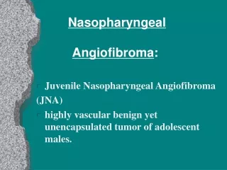
Nasopharyngeal Angiofibroma. Dr. Vishal Sharma. Definition. Benign tumor of nasopharynx (?), locally invasive, extremely vascular & occurs in adolescent males. Hamartomatous nidus of vascular tissue, dependent on testosterone. Synonyms: nasopharyngeal fibroma,

E N D
Nasopharyngeal Angiofibroma Dr. Vishal Sharma
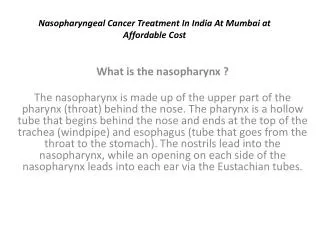
Definition • Benign tumor of nasopharynx (?), locally invasive, extremely vascular & occurs in adolescent males. • Hamartomatous nidus of vascular tissue, dependent on testosterone. • Synonyms:nasopharyngeal fibroma, angiofibroma
Site of origin Arises in posterior nasal cavity, near superior border of sphenopalatine foramen
Pathology Gross: Sessile, bi-lobed, rubbery, red-pink or gray in colour. Histology:Encapsulated, composed of vascular tissue & fibrous stroma. Vessels are thin-walled, lack elastic fibers & smooth muscle (this leads to uncontrolled bleeding).
Spread Anterior:Nasal cavity + paranasal sinus Posterior:Nasopharynx Lateral:goes to Pterygopalatine fossa 1. Infratemporal fossa cheek 2. Inferior orbital fissure orbit
Spread Superior:1. Sphenoid sinus Middle cranial fossa Cavernous sinus Optic chiasma Pituitary fossa 2. Skull base Middle cranial fossa
Symptoms 1. Nasal obstruction(80-90%)with denasal speech (rhinolalia clausa) 2. Epistaxis (50-60%): Persistent, Painless, Profuse, Paroxysmal, Unprovoked 3. Headache (25%) 4. Facial swelling (20%): cheek & palatal swelling
Signs 1. Nasal or Nasopharyngeal mass (80%) 2. Frog-face deformity: proptosis + nasal bridge broadening 3. Otitis media with effusion: due to E.T. blockage 4. Trismus: involvement of pterygoid muscle 5. Involvement of II, III, IV, VI cranial nerve
C.T. scan P.N.S. with contrast • Extent of tumor • Anterior bowing of posterior maxillary wall (Miller Holman’s antral sign) • Tumor enhancement • Bone destruction
Other Investigations M.R.I.:for intra-cranial involvement Digital Subtraction Angiography (D.S.A.):a. extent of tumor b. tumour blush (due to increasedvascularity)c. feeding arteries for embolization Biopsy: contraindicated (profuse bleeding)
Differential diagnosis • Rhabdomyosarcoma • Antrochoanal polyp • Teratoma • Dermoid • Encephalocoele • Inverting papilloma • Squamous cell carcinoma
Staging Stage I: Tumor limited to nasal cavity or nasopharynx with no bony destruction Stage II:Tumor invading pterygopalatine fossa or paranasal sinusesStage III: Tumor invading infratemporal fossa or orbit or parasellar region Stage IV:Tumor invading cavernous sinus or optic chiasma or pituitary fossa
Pre-op reduction of tumor vascularity 1. Embolization of feeding arteries: with Gelfoam 2. Oestrogen therapy:Diethylstilbestrol (2.5 - 5 mg orally t.i.d. for 3 - 6 wk) 3. Testosterone receptor blocker:Flutamide 4. Pre-operative radiotherapy 5. Cryotherapy of tumor
Denker’s incision Caldwell Luc incision extended medially till midline
Surgical approaches 1. Trans-palatal approach (Wilson) small tumour in nasopharynx 2. Sublabial + Trans-palatal approach (Sardana) large tumour of nose + PNS + nasopharynx 3. Intranasal endoscopic approach small tumour in nose / PNS / nasopharynx
Surgical approaches 4. Transmaxillary approach via: Extended lateral rhinotomy incision Midfacial degloving incision Denker’s extended Caldwell-Luc incision Le Fort 1 osteotomy approach Done for extension into pterygopalatine fossa
Surgical approaches 5. Infratemporal fossa approach (Fisch) extension into infratemporal fossa 6. Anterior subcranial approach intracranial & orbital extension 7. Image-guided, endoscopic, laser-assisted removal (latest): small / medium size tumors
Proton Stereotactic Radiation Therapy (P.S.R.T.) Synonym:Gamma knife surgery Used for: 1. Intracranial extension 2. Recurrence after surgery • Single relatively high dose of radiation delivered precisely to a small area to kill tumorcells • Minimal injury to adjacent nerves & brain tissue

![Nasopharyngeal Cancer [6]](https://cdn1.slideserve.com/3354426/nasopharyngeal-cancer-6-dt.jpg)