Download
1 / 21
210 likes | 213 Vues
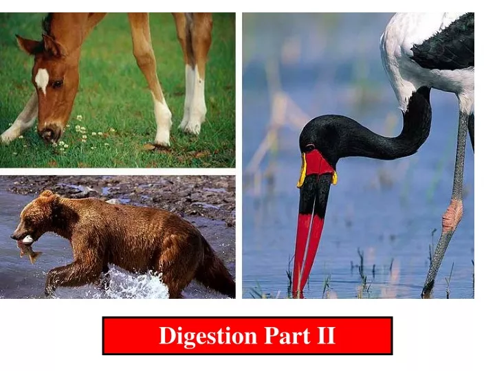
This article explores the process of human digestion from the mouth to the small intestine, focusing on the role of each organ and the enzymes involved. Learn about the functions of the mouth, esophagus, stomach, and small intestine in breaking down food and extracting nutrients.

E N D
Human Digestion Mouth- teeth used to mechanically break down food Tongue- moves food around, monitors texture and chemistry via chemoreceptors (taste buds) Taste of food stimulate saliva Saliva glands- produce a substance known as saliva. Saliva-95% H2O, ions, lubricating mucus, and amylase (breaks up starch)
Amylase enzyme works well in slightly alkaline solutions After food is chewed sufficiently, it is called a bolus and is swallowed down the pharynx which joins the nasal cavity Pharynx- anterior larynx- when raised closes trachea -posterior laryngopharynx When swallowing occurs the pharynx closes off the trachea to push food down the esophagus
Esophagus -upper is under voluntary control nervous system -lower is under involuntary control nervous system There are two layers of muscle around the esophagus. One is longitudinal and the other is circular. The two will squeeze together to form a peristaltic wave. A number of these waves are used to move the bolus down to the stomach. The digestive tract has three layers, which can be seen throughout the entire gastrointestinal tract.
1. Muscularis- outer layer A. serosa outer fiber layer B. circular and longit- udinal muscle (smooth, involuntary muscle) 2. Submucosa- connective tissue, blood vessels, nerves, lymph vessels and glands 3. Mucosa- inner layer epithelium connective tissue and lymph nodes
The bolus moves down the esophagus via peristalsis. The bolus enter the stomach via cardiac sphincter which is muscle that opens and closes the stomach. Stomach has the standard three layers with an additional layer of diagonal muscle in the muscularis; allows for peristalsis. The upper part of the stomach has pits with leading to glands. Parietal cells produce HCl acid. Mucus cells that produce mucus to protect the lining of the stomach. Chief cells secrete pepsinogen (precursor of pepsin). These produce gastric juices. Pepsin is an enzyme that starts to breakdown proteins.
Hydrochloric acid is used to destroy bacteria found on food, breakdown other materials like bone, eggshell and other things. The pH of the hydrochloric acid is around 2. The Cl- comes from salt and the H+ comes from water.
Once the bolus enters the stomach it distends; this distention causes increase motility and a release of a hormone gastrin. The release of gastrin stimulates the release of HCl and pepsinogen (precursor of pepsin); pepsinogen is made into an active enzyme pepsin by HCl and other pepsin Enzymes secreted by the stomach Pepsin- hydrolyzes protein to polypeptides Rennin- soluble milk protein- milk curd Gastric lipase- neutral fats- F.A. and glycerol and does contribute to lipid digestion (not all )
Peristalsis moves the bolus back and forth digesting it to chyme. Very acidic 1.5-2 pH; coating of mucus prevents acid from breaking down the tissue of the stomach. Finally the acidic chyme passes into the pyloric sphincter into the duodenum or small intestines. The small intestine (4.5 meters long/ 20 ft.) is kept in place by a membrane called mesentery. The first 25 cm is called the duodenum. The rest of the small intestine is divided up into the jejunum and the ileum. A major portion of digestion of carbohydrate, protein, lipid and nucleic acid digestion occurs there. Absorption of nutrients occurs also occurs there.
Acidic chyme passes into the duodenum which causes the secretion of two hormones (CCK and secretin). Both of these hormones causes stomach to stop peristalsis and HCl production. Cholecystokinin (CCK) is induced by the presence of fat It causes the release of pancreatic enzymes and the contraction of the gall bladder. Secretin is induced by the acid in the chyme. It causes the pancreas to release sodium bicarbonate to neutralize the acid in the chyme. The small intestine produces digestive enzymes. The pancreas also produces digestive enzymes that are put into the small intestine.
Pancreas-is an accessory organ that secretes a number of enzymes through the pancreatic duct. It also secretes sodium bicarbonate to neutralize acid. Enzymes secreted by the pancreas 1. trypsin- breaks protein into smaller polypeptide chains.
2. Chymotrypsin- breaks proteins into smaller polypeptide chains difference is where the enzymes cut the chain. Note-both are secreted a zymogens (pre-cursor enzymes) and then once in the small intestine they become activated. 3. Pancreatic lipase- Breaks down fats into fatty acids and glycerol 4. Pancreatic amylase- Breaks starch down to maltose 5. Nucleases- breaks down nucleic acids into nucleotides The pancreas also produces hormones insulin and glucagon. These regulate glucose levels in the blood
Liver- secrets bile which is stored in gallbladder. Then the bile moves from the gallbladder to the small intestines. • Bile emulsifies fats so that lipase can break fats down. • Small intestine- has glands which also secretes digestive enzymes. • Dipeptidases- dipeptides-> single amino acids • 2. Carboxyl peptidase removes the end amino acid off of protein chain at the carboxyl end. • 3. Amino peptidase removes the end amino acid off of a protein at the amine end • 4. Maltase- maltose-> 2glucose • 5. Lactase- lactose-> glucose and galactose • 6. Sucrase- sucrose-> glucose and fructose
The small intestine is like a “wrinkled-terry cloth towel” The large folds increase surface area. Each finger-like projection (like the ones found on the towel) is termed a villus. The villi are lined with epithelial cells. The epithelial membrane facing the lumen of the small intestine is also folded. These folds are called microvilli. All the folding increases the surface area. A villus inside has structure called a lacteal which is connected to a lymph vessel.
The lacteal is surrounded by a capillary bed. The venule takes blood from the intestines to the hepatic portal vein, which takes the blood to the liver. The epithelial cells absorb nutrients by different methods. Fatty acids and monoglycerides are absorbed by passive diffusion in the epithelial cell. Then are reassembled into triglycerides. These
Triglycerides combines with phospho-lipids, cholesterol and proteins to form a chylomicron, which is transported to the lacteal and not the capillary beds. The lacteal puts the fat into the lymphatic system, which will dump the fats into a major vein near the heart. All the other nutrients move from the epithelial cell to the capillary bed. How the other nutrients move into the epithelial cell from the lumen of the small intestines, varies. Fructose is transported by facilitated diffusion and then transferred to the capillary bed. Glucose, amino acids and vitamins are transported by active transport.
Large Intestine- six feet long and its purpose is H2O and mineral reabsorption allowing fecal matter to become a semi-solid to go out cecum, Parts in humans 1. ascending, 2. transverse, 3. descending 4.rectum- anal canal- anus When food leaves the small intestine it passes into the large intestine or colon. The main job of the colon is to remove water from the undigested material to produce feces. Note that a major part of fecal matter is dead bacteria that live in the digestive tract and not undigested food matter. The greater the plant matter in the diet, the less compact the fecal matter. THIS is a GOOD thing.