Download
1 / 56
560 likes | 571 Vues
Genetic Regulation. OVERVIEW OF GENETIC REGULATION Regulation of gene expression is an essential feature in maintaining the functional integrity of a cell.

E N D
OVERVIEW OF GENETIC REGULATION • Regulation of gene expression is an essential feature in maintaining the functional integrity of a cell. • Increasing or decreasing the expression of a gene can occur through a variety of mechanisms, but many of the important ones involve regulating the rate of transcription. • In addition to the basic transcription proteins, RNA polymerase, sigma (prokaryotes), and TFIID (eukaryotes), activator and repressor proteins help control the rate of the process. These regulatory proteins bind to specific DNA sequences associated with both prokaryotic and eukaryotic gene regions. • Other mechanisms are important, and, especially in eukaryotes, gene expression is controlled at multiple levels.
REGULATION OF PROKARYOTIC GENE EXPRESSION • Regulation of gene expression in prokaryotes usually involves either initiation or termination of transcription. In bacteria, genes are often organized into operons. • An operon is a set of structural genes coding for a group of proteins required for a particular metabolic function along with the regulatory region(s) that controls the expression of the structural genes.
The regulatory region is upstream (tothe 5' side) from the structural genes and coordinates their regulation. An operon usually produces a polycistronic mRNA that carries the information for synthesis of all of the enzymes encoded by the structural genes. Twoexamples of transcriptional control in prokaryotes are discussed: • Regulation by activator and repressor proteins in the lactose operon • Attenuation control in the histidine operon
The lactose (lac) Operon • The lactose operon is a portion of the bacterial chromosome that controls the synthesis of three enzymes involved in the metabolism of the sugar lactose. • Most bacteria carry out glycolysis, a pathway that allows glucose to be metabolized as a carbon and energy source. • If glucose is unavailable, they can metabolize alternative carbohydrates, but require proteins (enzymes) in addition to those in glycolysis to do so. • Lactose, a disaccharide of galactose and glucose, represents one alternative sugar, and the genes of the lactose operon encode the additional proteins required for its metabolism.
The cell expresses these genes only when lactose is available and glucose is not. The structural genes of the lactose operon include: • The Z gene, which encodes a β-galactosidase (a prokaryotic lactase) • The Y gene, which encodes a galactoside permease, the transport protein required for entry of lactose into the cell • The A gene, which encodes a thiogalactoside transacetylase enzyme that is not essential for lactose metabolism and whose function is uncertain
In addition, the i gene, which encodes the lac repressor protein, is also considered part of the operon although it is located at a distant site in the DNA. The i gene is constitutively expressed (not regulated); thus, copies of the lac repressor protein are always in the cell. • Two gene regulatory proteins control the expression of the lac operon: • The lac repressor (encoded by the i gene), which binds to a DNA sequence called the operator • A cAMP-dependent activator protein (product of crp gene), CAP, which binds to a DNA sequence called the CAP site
Glucose and lactose control the expression by different mechanisms: • Lactose (or allolactose) induces gene expression by preventing the repressor protein binding to the operator sequence. • Glucose represses gene expression by lowering the level of cAMP in the cell, thus preventing the cAMP-dependent activator binding to the CAP-site sequence.
Coordinated Control of the Lactose Operon by Glucose and Lactose • Full expression of the lactose operon requires that both mechanisms favor gene expression. • The repressor protein must not bind at the operator, and • The cAMP-dependent activator protein must bind to the CAP site. This in turn requires that • Lactose is present (prevents repressor binding) • Glucose is low (to allow cAMP to increase)
Although intermediate levels of gene expression may be possible in the cell, it is convenient to simplify the situation in the following way: • The only condition that allows high gene expression is: lactose present, glucose absent. • All other combinations of these sugars result in low-level expression.
Attenuation in the Histidine Operon • The histidine operon encodes the enzymes of the histidine biosynthetic pathway. It is advantageous to the cell to produce these enzymes when histidine is not available in the surroundings, but to turn off their synthesis when histidine is readily available. • The histidine operon and several other operons for amino acid biosynthesis (e.g., tryptophan, leucine, and phenylalanine) are regulated by premature termination of transcription, a process known as attenuation.
Attenuation depends on the fact that transcription and translation occur simultaneously in bacteria. • In this process, transcription is constitutively initiated, and the 5' untranslated region (UTR) of the mRNA is followed by the coding region for a short (non-functional) peptide (the leader peptide). • If histidine is available in the growth medium, transcription is terminated before RNA polymerase reaches the structural genes of the operon • This form of regulation is dependent on the speed of the ribosome and the formation of two alternative secondary structures in the mRNA molecule.
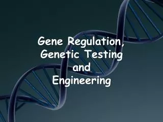
Attenuation Control of Transcription in the Histidine Operon
At High Levels of Histidine • As soon as the Shine-Dalgarno sequence associated with the leader peptide coding region appears in the 5' UTR of the mRNA, a ribosome binds and begins translating the message. • The ribosome can move quickly because it can easily find histidine to incorporate when it encounters histidine codons in the mRNA. • This allows the message to fold into a rho-independent terminator of transcription (stem and loop + poly-U). • RNA polymerase stops transcription before it reaches the structural genes, and no enzymes are produced.
At Low Levels of Histidine • A ribosome begins to synthesize the leader peptide, but stalls at the histidine codons because it cannot readily find histidine. • Because the ribosome is covering up a different part of the mRNA, the message will not fold into the correct terminator structure, and RNA polymerase continues transcription through the structural genes of the operon. • Translation of the message produces all the enzymes of the histidine biosynthetic pathway. Note: Attenuation is not used as a regulatory mechanism in eukaryotes, because transcription and translation are independent events and occur in different subcellular locations.
REGULATION OF EUKARYOTIC GENE EXPRESSION • In eukaryotic cells, DNA is packaged in chromatin structures, and gene expression typically requires activation to occur. Chromatin-modifying activities include: • Histone acetylases (favor gene expression) and deacetylases (favor inactive chromatin) • Scaffolding proteins that condense regions of the chromatin (favor inactive chromatin) • DNA methylating enzymes (favor inactive chromatin)
Activator proteins (and a few repressors) are important in eukaryotes, as they are in prokaryotes. • The DNA sequences to which activator proteins bind in eukaryotic DNA are called response elements. • A few response elements are located within the promoter region (upstream promoter elements [UPE]), but most are outside the promoter and often clustered to form an enhancer region that allows control of gene expression by multiple signals.
Upstream Promoter Elements • Only the proximity of the upstream promoter element to the -25 sequence distinguishes it from an enhancer. Proximal promoter elements include: • A CCAAT box (around -75) that binds a transcription factor NF-l (CTF) • A GC-rich sequence that binds a general transcription factor SP-l
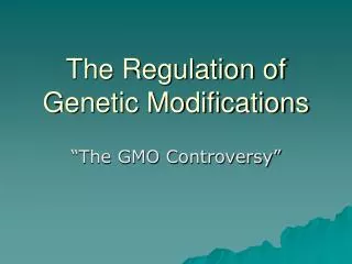
Enhancers • Enhancers in the DNA are binding sites for activator proteins. Enhancers have the following characteristics: • They may be up to 1000 base pairs away from the gene. • They may be located upstream, downstream, or within an intron of the gene they control. • The orientation of the enhancer sequence with respect to the gene is not important. • Enhancers can appear to act in a tissue-specific manner if the DNA-binding proteins that interact with them are present only in certain tissues. • Enhancers may be brought close to the basal promoter region in space by bending of the DNA molecule.
Stimulation of Transcription by an Enhancer and Its Associated Transcription Factors
Similar sequences that bind repressor proteins in eukaryotes are called silencers. There are fewer examples of these sequences known, and the mechanisms through which they act are not clear. Note • The Ig heavy chain locus has an enhancer in the large intron separating the coding regions for the variable domain from the coding regions for the constant domains Note Cis and Trans Regulatory Elements • The DNA regulatory base sequences (e.g., promoters, enhancers, response elements, and UPEs) in the vicinity of genes that serve as binding sites for proteins are often called "cis" regulators. • Transcription factors (and the genes that code for them) are called "trans" regulators. Trans regulatory protein can diffuse through the cell to their point of action.
Transcription Factors • The activator proteins that bind response elements are often referred to as transcription factors. Typically, transcription factors contain at least two recognizable domains, a DNA-binding domain and an activation domain. 1. The DNA-binding domain binds to a specific nucleotide sequence in the promoter or response element. Several types of DNA-binding domain motifs have been characterized and have been used to define certain families of transcription factors. Some common DNA-binding domains include: • Zinc fingers (steroid hormone receptors) • Leucine zippers (cAMP-dependent transcription factor) • Helix-loop-helix • Helix-turn-helix (homeodomain proteins encoded by homeotic/homeobox genes)
2. The activation domain allows the transcription factor to: a) Bind to other transcription factors b) Interact with RNA polymerase II to stabilize the formation of the initiation complex c) Recruit chromatin-modifying proteins such as histone acetylases or deacetylases • Two types can be distinguished, general transcription factors and specific transcription factors. Examples are listed in Table 1-5-1.
Each “finger” binds small stretch of DNA • Additive affect of each finger adds specificity and binding affinity to entire domain Mouse Zif268
Leucine zipper: Have basic amino acids with + charge (bind DNA) • E.g., cAMP dependent binding protein
Helix-Loop-Helix: Have basic helix with + charge (bind DNA) • Best characterized in mammalian MyoD protein (involved in muscle cell development)
Helix-turn-helix DNA-binding protein • Extra N-terminal domain that inserts into minor groove • First eukaryotic examples found fruit fly “homeotic” genes • mutations in these genes cause body-part changes
General Transcription Factors • In eukaryotes, general transcription factors must bind to the promoter to allow RNA polymerase II to bind and form the initiation complex at the start site for transcription. • General transcription factors are common to most genes. The general transcription factor TFIID (the TATA factor) must bind to the TATA box before RNA polymerase II can bind. Other examples include SP-l and NF-l that modulate basal transcription of many genes.
Specific Transcription Factors • Specific transcription factors bind to enhancer regions or, in a few cases, to silencers and modulate the formation of the initiation complex, thus regulating the rate of initiation of transcription. • Each gene contains a variety of enhancer or silencer sequences in its regulatory region. The exact combination of specific transcription factors available (and active) in a particular cell at a particular time determines which genes will be transcribed at what rates. • Because specific transcription factors are proteins, their expression can be cell-type specific. Additionally, hormones may regulate the activity of some specific transcription factors. Examples include steroid receptors and the CREB protein.
Peroxisome proliferator-activated receptors (PPARs) are transcription factors that bind to DNA response elements (PPREs) and control multiple aspects of lipid metabolism. Individual members of this family of zinc-finger proteins are activated by a variety of natural and xenobiotic ligands, including: • Fatty acids • Prostaglandin derivatives • Fibrates • Thiazolidinediones • The improvement in insulin resistance seen with thiazolidinediones is thought to be mediated through their interaction with PPARγ. Clofibrate binds PPARα affecting different aspects of lipid metabolism than the thiazolidinediones.
An example of how response elements affect metabolism can be seen in the pathway of gluconeogenesis. • Gluconeogenesis is a hepatic pathway whose major function is to maintain adequate glucose in the blood for tissues like the nerves (brain) and red blood cells during fasting. It also provides glucose during periods of stress. • Hormones that activate the pathway include: • Glucagon secreted in response to hypoglycemia and functioning via a membrane associated receptor that increases cAMP concentration • Cortisol secreted in response to stress, is permissive for glucagon in hypoglycemia and acts through an intracellular receptor, which, like other steroid receptors, is a zinc-finger DNA binding protein.
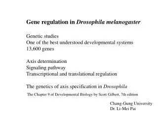
Control of Gluconeogenesis by Response Elements Cortisol and Glucagon Stimulate Gluconeogenesis Through Enhancer Mechanisms
Phosphoenolpyruvate carboxykinase (PEPCK) catalyzes a critical reaction in gluconeogenesis, which under many conditions is the rate-limiting step in the pathway. • A cAMP response element (CRE) and a glucocorticoid response element (GRE) are each located upstream from the transcription start site.
Cortisol induces PEPCK gene expression by the following sequence: • Cortisol diffuses into the hepatocyte • Binds to its receptor. • The complex enters the nucleus, and • Binds (through the zinc fingers) to the glucocorticoid response element (GRE) associated with the PEPCK gene, which • Increases gene expression. • PEPCK concentration increases in the cell. • The rate of gluconeogenesis increases.
Glucagon induces PEPCK gene expression by the following sequence: • Glucagon binds to a receptor in the cell membrane. • cAMP concentration increases. • Protein kinase A becomes active, and then • Phosphorylates and activates CREB. • Activated CREB enters the nucleus and binds to the CRE associated with the PEPCK gene, which • Increases gene expression. • PEPCK concentration increases in the cell. • The rate of gluconeogenesis increases. • These effects ofCREB and the cortisol-receptor complex are not entirely independent ofeach other. Each contributes, along with several other transcription factors, to assembling a complex ofactivator proteins that ultimately determine the level ofPEPCK gene expression.
Control of Cell Differentiation by Homeodomain Proteins During Development In Utero • Sequential and coordinated gene expression is necessary for proper tissue and cell differentiation during embryonic life. • Groups of regulatory proteins called homeodomain proteins are major factors in controlling this embryonic gene expression. • Each regulatory protein is responsible for activating a different set of genes at the proper time in development. • The regulatory proteins themselves are encoded by genes called homeobox (HOX) or homeotic genes. • Another closely related set of genes is the PAX (paired-box) genes. • Mutations in HOX or PAX genes might be expected to produce developmental errors. Klein-Waardenburg syndrome (WS-III) is one such developmental disorder resulting from a mutation in a PAX gene.
Clinical Correlate Klein-Waardenburg Syndrome • All of the tissues affected in Klein-Waardenburg syndrome are derived from embryonic tissue in which PAX-3 is expressed. Symptoms include: • Dystopia canthorum (lateral displacement of the inner corner of the eye) • Pigmentary abnormalities (frontal white blaze of hair, patchy hypopigmentation of the skin, heterochromia irides) • Congenital deafness • Limb abnormalities
Co-Expression of Genes • Most eukaryotic cells are diploid, each chromosome being present in two homologous copies. The alleles of a gene on the two homologous chromosomes are usually co-expressed. • In a person heterozygous for the alleles of a particular gene, for example a carrier of sickle cell trait, two different versions of the protein will be present in cells that express the gene. • In the person heterozygous for the normal and sickle alleles, about 50% of the β-globin chains will contain glutamate and 50% valine at the variable position (specified by codon 6).
Major exceptions to this rule of codominant expression include genes: • On the Barr body (inactivated X chromosome) in women • In the immunoglobulin heavy and light chain loci (ensuring that one B cell makes only one specificity of antibody) • In the T-cell receptor loci • Genomic Imprinting
Bridge to Medical Genetics Genetic Imprinting in Prader-Willi Syndrome • Genetic imprinting of a few gene regions results in mono-alleleic expression. • In some cases this imprinting is according to the parent of origin. The gene(s) involved in Prader-Willi syndrome is on chromosome 15 and is imprinted, so that it is normally expressed only from the paternal, not the maternal, chromosome. • In such a case, if one inherits a paternal chromosome in which this region has been deleted, Prader-Willi syndrome results. It can also result from uniparental (maternal) disomy of chromosome 15. • Symptoms of Prader-Willi include: • Childhood obesity and hyperphagia • Hypogonadotrophic hypogonadism • Small hands and feet • Mental retardation • Hypotonia
Angelman Syndrome • Characterized by unusual facial appearance, short stature, severe mental retardation, spasticity, and seizures • Have genetic info in 15q12 (15q11-q13) derived only from father
Other Mechanisms for Controlling Gene Expression in Eukaryotes • Table 1-5-2 summarizes some of the mechanisms that control gene expression in eukaryotic cells.