Download
1 / 15
150 likes | 198 Vues
Sameer Ahmed 9/25/2013. Cummings Ch 127: T-Bone and Ear Anatomy. The temporal bone consists of four embryologically distinct components: Squamous, mastoid, petrous, and tympanic parts

E N D
Sameer Ahmed 9/25/2013 Cummings Ch 127: T-Bone and Ear Anatomy
The temporal bone consists of four embryologically distinct components: • Squamous, mastoid, petrous, and tympanic parts • A horizontal ridge known as the temporal line is formed along the most inferior insertion by the temporalis muscle • Aligned with the zygomatic process, • Surface landmark that estimates the location of the middle fossa floor (Tegmen) • “The middle fossa dural plate (MFD) is located on average 5 mm above the LTI (linea tempralis inferior)” • “This study confirms that the mastoid antrum is located 15 mm deep to the lateral surface of the mastoid bone”
Facial Nerve Course • Cisternal segment/intracranial segment • Brainstem to IAC --> 16 -24 mm • Meatal segment → 8mm • Porus acoustics: Medial IAC • Fundus: Lateral IAC • Labyrinthine segment → 5mm • Fundus to geniculate ganglion(1st genu) • Tympanic segment → 8-11mm • Geniculate to 2nd genu • Courses above oval window; dehisc 50% of time • 2nd genu is just anteroinferior to the horizontal SCC • Vertical segment → 10-14mm • 2nd genu to stylomastoid foramen
At the fundus (lateral IAC) • Bill's Bar separates CN 7 anteriorly from SVN • “Jail Bar” • Falciform cr. separates CN 7 from cochlear n.; SVN from IVN • Anatomic relationship changes closer to the brainstem
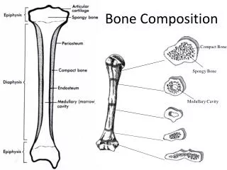
Jugular foramen: • Pars nervosa: CN X, CN XI, Arnold's nerve, jugular bulb, and posterior meningeal branch of ascending pharyngeal artery • Pars venosa: CN IX, Jacobson nerve, and venous return from inferior petrosal sinus • Inferior limit for a translab approach to IAC • Cochlear aqueduct • The cochlear aqueduct eventually opens into the scala tympani at the cochlear base • Keel: Ridge of bone between Jugular bulb and ICA
EAC: • Lateral cartilaginous: 1/3rd • Lateral canal skin thicker, more sebaceous units • Medial bony: 2/3rd • Think skin; continuous with TM epithelium • Bony-cartilaginous junction in EAC • Site of granulation tissue in malignant OE • Routes of tumor/infxn spread • Foramen of Hushke • Incomplete ossification of bony anterior EAC; medial • Fissures of Santorini • Defects in cartilaginous EAC
1st and 2nd branchial arches → external ear • 6 Hillocks of His • 1st branchial arch: 1-3 hillocks (tragus, superior helix) • 2nd branchial arch: 4-6 hillocks (antihelix, antitragus, lobule, and inferior helix) • External ear blood supply • Posterior auricular & Superficial temporal art.
Tympanic Membrane • Outer layer: epidermal/squamous (ectoderm) • Middle layer: fibrous (mesoderm) • Can be subdivided into radial outer and circular inner • Inner layer: mucosal (endoderm) • Fibrous annulus: thickened pars tensa forming a fibrous outler ring for the attachement to the T-Bone; lies within tympanic sulcus except superiorly where it is deficient at the Notch of Rivinus • Pars flaccida = Shrapnell's Membrane
Eustachain Tube • Medial 1/3rd → Bony • Lateral 2/3rd → Cartilaginous • Collapsed at rest • Tensor veli palatini intermittenly enlarges ET during yawning or swallowing • Bony cartilaginous jnxn of ET is the narrowest portion of the ET • Carotid artery is just medial to ET
Middle Ear • Relative to tympanic annulus • Epitympanum: above annulus • Mesotympanum: confined by the annulus • Hypotympanum: below annulus • Mestoympanum • Anterior limit: ET • Posterior limit: Facial nerve • Medial wall: Cochlear promontory • Postero-superior: Oval window • Postero-inferior: Round window • Sinus tympani: posterior to both windows and medial to vertical division of FN • Pyramidal eminence: anterior to 2nd genu
Hypotympanum • Limited inferiorly by the jugular bulb • Epitymapnum • Superior-medial wall of bony EAC (scutum) forms the lateral wall of the epitympanum • Epitympanum divided into 3 spaces: • Prussak's space, just medial to pars flaccida and lateral to the head and neck of the malleus; • The compartment anterior to the malleus • The posterior compartment, which communicates with the antrum • Attic Cholesteatomas can spread postero-superiorly into the antrum or postero-inferiorly into posterior mesotympanum
Ossicles • Malleus • Manubrium (handle; tip is the umbo) • Lateral/short process • Anterior process • Head • Neck • Tensor tympani attaches to malleus neck and manubrium by a tendon originating from the cochleariform process
Incus • Body • Short process • Long process • Lenticular process • Diarthrodial joints (for malleus-incus and incus-stapes connections)
Stapes • Head (capitulum) • Anterior and posterior crus • Footplate (base) • Stapes footplate attaches to bony margin of oval window via an annular ligament (syndesmosis joint)