Download

1 / 49
510 likes | 704 Vues
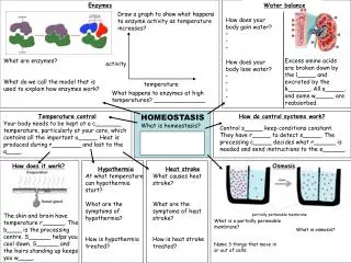
Alterations In Homeostasis. Shock. Homeostasis. What is homeostasis?????

E N D
Homeostasis • What is homeostasis????? • Homeostasis is an (ideal or virtual) state of equilibrium, in which all body systems are working and interacting in an appropriate way to fulfill all the needs of the person and/or the body. When homeostasis is interrupted (e.g. by response to a stressor), the body tries to restore it by adjusting one or more physiological processes. This stress-adaption mechanism includes activation of the Hypothalamic-Pitauitary-Andrenal Axis (HPA Axis) with the autonomous nervous system and endocrine reactions of the body. • Severe stressors or long lasting adjustment demands can cause severe imbalance of this steady state. This might cause not only psychological distress but also psycho-somatic disorders.
Shock • Can occur when any part of the cardiovascular system does not function properly for any reason • Begins with abnormal cellular metabolism that occurs when too little oxygen is delivered to tissues • Shock is a condition in which a systemic decrease in perfusion to tissue and organs leads to poor gas and nutrient exchange. Delays in recognition and treatment can lead to irreversible shock, multisystem organ failure, and death. • Types of shock and their causes vary because shock is a manifestation of a pathologic condition rather than a disease state. More then one type of shock can be present at one timie
Process of Shock • Initial stage (early shock) • Nonprogressive stage (compensatory stage) • Progressive stage (intermediate stage) • Refractory stage (irreversible stage)
Initial stage (early shock) Reversible • Pathophysiology
Nonprogressive Stage (Compensatory) • Pathophysiology
Progressive Stage (Intermediate Stage) • Pathophysiology
Refractory stage (Irreversible Stage) • Pathophysiology
Multiple Organ Dysfunction Syndrome • Pathophysiology
Effects of Shock on Body Systems • Cardiovascular System
Effects of Shock on Body Systems • Respiratory System
Effects of Shock on Body Systems • GI System/Renal
Effects of Shock on Body Systems • Neurologic System
Effects of Shock on Body Systems • Skin, Temperature, and Thirst
CVP Normal CVP: 0-6mm Hg Increased CVP: • Aggressive fluid resuscitation • right-sided heartfailure with venoconstriction • renal failure • tricuspid or pulmonic valvular disorders • right ventricular infarction • COPD • pulmonary embolis • pulmonary hypertension. What does the patient look like? dyspnea, crackles, distended neck veins
CVP Decreased CVP • Hypovolemia-relative or actual • Hemmorhage • Vasodilitation • Diuretics • Fliud shifts-sepsis What does the patient look like? Tachycardia CVP will fall before the patient becomes hypotensive.
Complication of CVP Catheters • Infection • Thrombosis • Hemorrhage • Arrhythmia • Pneumothorax • Cardiac Tamponade
Pulmonary Artery Catheter • Used to continuously monitor right atrium (RA) and pulmonary artery (PA) pressures. • Swan-Ganz Catheter • Insertion sites: subclavian, internal or external jugular, femoral, brachial.
Left Heart Preload/PCWP PAOP=PCWP=LVEDP=left heart preload Measures filling pressures in the left heart Normal: 5-12mmHg PAD=PCWP in the absence of pulmonary hypertension
PCWP PCWP normally correlates with volume Low PCWP indicates hypovolemia (Volumeexpanders, packed RBC) High PCWP indicates hypervolemia (diuretics, venodilators)
Cardiac Output/Cardiac Index Normals: CO: 4-8L/min; CI:2.5-4.0L/min/m2 What can cause Low CO/CI: • HR: Fast or slow • Preload: decreased from diuresis, dehydration, fluid shifts, hypovolemia, vasodilitation • Afterload: Increased from vasoconstriction secondary to HTN, compensatory vasoconstriction • Contractility: Decreased from MI, HF, cardiomyopathy, cardiogenic shock, cardiac tamponade
Cardiac Output/Cardiac Index High CO/CI: • Anxiety • Compensatory response in pulmonary edema • Increased metabolic states (fever, hyperthyroid) • sepsis
Right Heart Afterload • Right heart afterload=pulmonary vascular resistance (PVR) • Normal: 50-250 dynes/sec/cm2 • Increased in acute lung injury
Left Heart Afterload • Left heart afterload=systemic vascular resistance (SVR) • Normal: 800-1200 dynes/sec/cm2
Increased Afterload (SVR) • Use of vasopressors • Aortic stanosis • hypothermia • hypertension
Increased Afterload (SVR) • Treatment • Vasodilators (nipride, NTG) • Ace Inhibitors (captopril, enalapril) • Calcium channel blockers (verapamil nifedipine)
Decreased Afterload (SVR) • Sepsis/septic shock • Anaphylactic Shock • Neurogenic Shock • Treatment • Levophed • Neosynephrine • Dopamine • Vasopressin
Indication for an A-line • Critically ill patients with intra-aortic balloon pumps • Monitor the effects of potent vasodilators and vasopressors • Frequent ABG testing • Morbid obesity • Burn patients
Nursing Process • Assessment • Health History • Physical Examination • Nursing Diagnosis • Plan • Implantation • Evaluation