Download
1 / 10
120 likes | 146 Vues
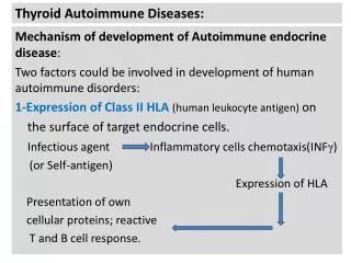
Autoimmune Diseases. Occasionally the immune system loses the ability to recognize the body’s own cells. The immune system then begins destroying the body’s own organs or tissues. This is called and autoimmune disease.

E N D
Autoimmune Diseases • Occasionally the immune system loses the ability to recognize the body’s own cells. The immune system then begins destroying the body’s own organs or tissues. This is called and autoimmune disease. • It is not clear why this happens in all cases. 4 types of autoimmune diseases: • 1. Autoimmunity: • Antibodies made by the body that attack body tissues or organs. • May be due to antibodies made in response to foreign antigens, on microorganisms, with a similar sequence to the body cells’ proteins. The immune system is unable to distinguish between the two proteins because they are so similar.
2. Cytotoxic: • Mistaken antibody reactions to cell-surface proteins. • Myasthenia gravis: muscles become progressively weaker due to antibodies that bind to acetylcholine receptors on nerves that reach the muscles • Eventually muscles fail because do not receive the proper chemical signals. • Usually results in respiratory arrest and death • It is not known why the body makes antibodies to a chemical receptor like this. • 3. Immune Complex: • Just like with the hypersensitive immune complex disease, this type of autoimmune disease is caused by clusters of antibody/antigen deposited in the body. • Rheumatoid arthritis: immune complexes deposited in joints. • Causes chronic inflammation and leads to severe damage to cartilage and joint due to the immune system’s efforts to get rid of the immune complex deposits.
4. Cell-mediated: • Insulin-dependent mellitus: immunological destruction of insulin-secreting cells of the pancreas. • T cell dysfunction is implicated in disease.
Phenotypic Methods: manifest characteristics. • Microscopic Examination: • Gram Stain, Acid Fast Stain, Fluorescence Antibody Test, and Antigen Testing are all quick ways to diagnose common infections. • Cultivation: • Selective media and Differential media • Motility, Oxygen requirements, cultural appearance
Biochemical Testing • Carbohydrate fermentation (lactose, sucrose, glucose, etc.) • Enzymes produced (catalase, oxidase, bile esculin, etc.)
Genotypic Methods: what the genes say. • DNA Analysis • Hybridization: small fragments of DNA (probes) of known sequence attach to unknown DNA. When they attach, the organisms is identified! • rRNA Analysis • Sequence of bases in ribosomes. • PCR • Used to sequence a known gene for a particular organism. If the gene is replicated then that organism is known to be present in the sample. • GC base content • Each genus has differing amounts of A/T to C/G ratios within DNA. This technique is more useful in determining ancestral relationships rather than identifying a microorgansim.
Immunological Methods • Antigen-Antibody Rxns: • Agglutination and Precipitation Rxns: • Whole cell of the sample organism combined with Ab for a suspected organism. This test is conducted on a slide. If the Antibody (Ab) matches the organism in the sample, the antibodies will bind to the organism and visible clumps will form in the solution. • Ag with Ab (saline agar plate). This is the same concept as the one above however it is conducted on an agar plate and the antigen/antibody reaction is seen within the agar. • Western Blot • Used for HIV confirmation • The western blot is a combination of gel electrophoresis, to separate specific proteins in a sample, and an antigen/antibody reaction. • Once the proteins (possible HIV antibodies) are separated on the gel, HIV antigens are added to see if they will bind to any of the protein bands from the gel. If any of the antigens bind, then that sample is positive for antibodies for HIV.
ELISA (Enzyme-Linked Immunosorbent Assay) Used for HIV screen tests. • See Figure 17.16 • 1. The known antigen, anti-HIV, is adsorbed to the well. • 2. The serum samples from the patient are then place in the well. • If the serum contains antibodies for HIV, they will bind to the antigen. • 3. An indicator molecule is put into the well that will bind to the HIV antibody if it is still present. • When it binds to the antibody it catalizes a reaction that releases a dye.
I didn’t go into a lot of detail on most of these techniques because I felt that most of them were pretty self explanatory. • If you more help understanding them please email me or talk to me about it in class on Friday.