Download
1 / 1
10 likes | 113 Vues
Importance of Brain Reward Regions in Adults with Acquired Structural Hypothalamic Damage: A Functional Neuroimaging Study

E N D
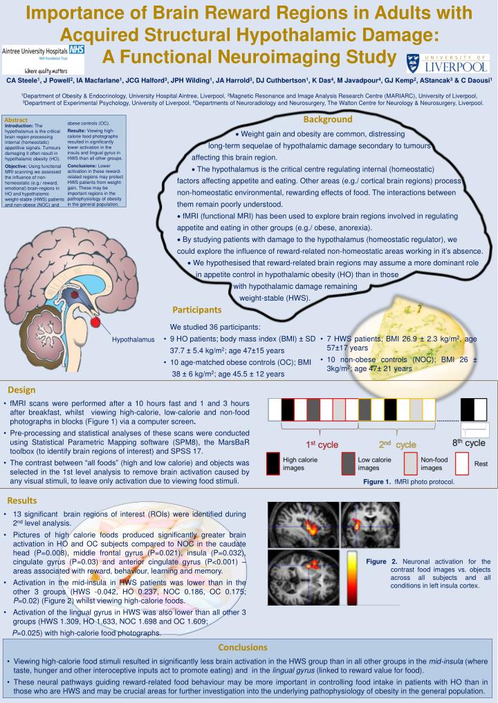
Importance of Brain Reward Regions in Adults with Acquired Structural Hypothalamic Damage: A Functional Neuroimaging Study CA Steele1, J Powell2, IA Macfarlane1, JCG Halford3, JPH Wilding1, JA Harrold3, DJ Cuthbertson1, K Das4, M Javadpour4, GJ Kemp2, AStancak3 & C Daousi1 1Department of Obesity & Endocrinology, University Hospital Aintree, Liverpool, 2Magnetic Resonance and Image Analysis Research Centre (MARIARC), University of Liverpool, 3Department of Experimental Psychology, University of Liverpool, 4Departments of Neuroradiology and Neurosurgery, The Walton Centre for Neurology & Neurosurgery, Liverpool. Abstract Introduction: The hypothalamus is the critical brain region processing internal (homeostatic) appetitive signals. Tumours damaging it often result in hypothalamic obesity (HO). Objective: Using functional MRI scanning we assessed the influence of non-homeostatic (e.g./ reward, emotional) brain-regions in HO and hypothalamic weight-stable (HWS) patients and non-obese (NOC) and obese controls (OC). Results: Viewing high- calorie food photographs resulted in significantly lower activation in the insula and lingual gyrus in HWS than all other groups. Conclusions: Lower activation in these reward- related regions may protect HWS patients from weight- gain. These may be important regions in the pathophysiology of obesity in the general population. Hypothalamus Background Weight gain and obesity are common, distressing long-term sequelae of hypothalamic damage secondary to tumours affecting this brain region. The hypothalamus is the critical centre regulating internal (homeostatic) factors affecting appetite and eating. Other areas (e.g./ cortical brain regions) process non-homeostatic environmental, rewarding effects of food. The interactions between them remain poorly understood. fMRI (functional MRI) has been used to explore brain regions involved in regulating appetite and eating in other groups (e.g./ obese, anorexia). By studying patients with damage to the hypothalamus (homeostatic regulator), we could explore the influence of reward-related non-homeostatic areas working in it’s absence. We hypothesised that reward-related brain regions may assume a more dominant role in appetite control in hypothalamic obesity (HO) than in those with hypothalamic damage remaining weight-stable (HWS). Participants • We studied 36 participants: • 9 HO patients; body mass index (BMI) ± SD • 37.7 ± 5.4 kg/m2; age 47±15 years • 10 age-matched obese controls (OC); BMI • 38 ± 6 kg/m2; age 45.5 ± 12 years • 7 HWS patients; BMI 26.9 ± 2.3 kg/m2, age 57±17 years • 10 non-obese controls (NOC); BMI 26 ± 3kg/m2; age 47± 21 years Design • fMRI scans were performed after a 10 hours fast and 1 and 3 hours after breakfast, whilst viewing high-calorie, low-calorie and non-food photographs in blocks (Figure 1) via a computer screen. • Pre-processing and statistical analyses of these scans were conducted using Statistical Parametric Mapping software (SPM8), the MarsBaR toolbox (to identify brain regions of interest) and SPSS 17. • The contrast between “all foods” (high and low calorie) and objects was selected in the 1st level analysis to remove brain activation caused by any visual stimuli, to leave only activation due to viewing food stimuli. Figure 1. fMRI photo protocol. Results • Figure 2. Neuronal activation for the contrast food images vs. objects across all subjects and all conditions in left insula cortex. • 13 significant brain regions of interest (ROIs) were identified during 2nd level analysis. • Pictures of high calorie foods produced significantly greater brain activation in HO and OC subjects compared to NOC in the caudate head (P=0.008), middle frontal gyrus (P=0.021), insula (P=0.032), cingulate gyrus (P=0.03) and anterior cingulate gyrus (P<0.001) – areas associated with reward, behaviour, learning and memory. • Activation in themid-insula in HWS patients was lower than in the other 3 groups (HWS -0.042, HO 0.237, NOC 0.186, OC 0.175; P=0.02) (Figure 2)whilst viewing high-calorie foods. • Activation of the lingual gyrus in HWS was also lower than all other 3 groups (HWS 1.309, HO 1.633, NOC 1.698 and OC 1.609; • P=0.025) with high-calorie food photographs. Conclusions • Viewing high-calorie food stimuli resulted in significantly less brain activation in the HWS group than in all other groups in the mid-insula (where taste, hunger and other interoceptive inputs act to promote eating) and in the lingual gyrus (linked to reward value for food). • These neural pathways guiding reward-related food behaviour may be more important in controlling food intake in patients with HO than in those who are HWS and may be crucial areas for further investigation into the underlying pathophysiology of obesity in the general population.