Download
1 / 83
910 likes | 1.27k Vues
Thank You. Introduction By Zeinab Salah . The History of Apoptosis. Cell death was first discovered by a German scientist. The term programmed cell death was introduced.

E N D
Introduction By Zeinab Salah
The History of Apoptosis • Cell death was first discovered by a German scientist. • The term programmed cell death was introduced. • Term apoptosis coined by Greek scientists ( Kerr, Wyllie ,Currie and James Cormack). • The word "apoptosis" (Greek spelling of apoptosis) is used in Greek to describe the "dropping off" or "falling off" of leaves from trees. • Apoptosis genes identified , ( Bcl 2 , p53 , ).
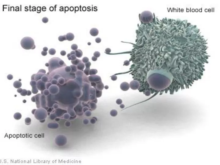
Apoptosis :- Apoptosis (programmed cell death) or death comes from the cell Is a mode of cell death that occurs under normal physiological conditions and the cell is an active participant in its own demise (cellular suicide). It is one of the main types of programmed cell death (PCD) occurs in multicellular organisms and involves a series of biochemical events leading to specific cell morphology characteristics and ultimately death of cell.
Apoptosis is caused by activation of intracellular proteases, known as caspases, that are responsible directly or indirectly for the morphologic and biochemical events that characterize the apoptotic cell , these morphological changes, including:- 1.Cell shrinkage and chromatin condensation . 2.Blebbing of the cell membrane . 3.Nuclear fragmentation . 4.Formation of Apoptotic bodies . 5. Phagocytosis without inflammatory response
Scanning electron micrographs of normal and apoptotic thymocytes
In contrast to necrosis, which is a form of traumatic cell death that results from acute cellular injury. • It is a death of a group of cells often associated with extensive tissue damage resulting in an intense inflammatory response.
Physiological Roles of Apoptosis:- • Programmed cell death is as needed for proper development :- • Embryonic Development :- During development for removal of excess cells during embryogenesis , such as The formation of the fingers and toes of the fetus requires the removal, by apoptosis, of the tissue between them . syndactylism
When a tadpole changes into a frog, the cells in the tadpole tail are induced to undergo apoptosis, so the tail is lost, which is not needed in the frog. • Apoptosis during the metamorphosis of a tadpole into a frog:-
Neurological development :- • The formation of the proper connections (synapses) between neurons in the brain requires that extra cells be eliminated by apoptosis. • Involution of tissue:- • For example, endometrial breakdown during the menstrual cycle and regression of lactating breast tissue after weaning involve apoptosis.
Maintenance of homeostasis :- • To maintain cell population in constant number tissues with high turnover of cells, such as skin.
Immune System:- • Death of cells that have served their use full purpose, such as neutrophils in acute inflammatory response , and activated lymphocytes at the end of an immune response . • Elimination of potentially harmful self-reactive lymphocytes. • .
Pathological Roles of Apoptosis:- Death by apoptosis is responsible for loss of cells in a variety of pathologic States :- • Cell with DNA damage increase the expression of genes considered as a potent inducer of apoptosis. So elimination of the cell may be better than risking mutations in the damaged DNA .
Accumulation of mis-folded proteins • Improperly folded proteins may arise because of mutation in the genes encoding these proteins .Excessive accumulation of these proteins in ER lead to condition called ‘’ ER stress’’ which terminate in apoptotic death of the cell. • Cell death induced by cytotoxic T lymphocytes against viruses and tumors.
Apoptosis and the cell cycle • The cell cycle is divided into four phases:-
Are proliferation and apoptosis linked ? • Cell proliferation, differentiation and death are fundamental processes in multicellular organisms, and several lines of evidence link apoptosis to proliferation. • Firstly, uncontrolled proliferation can be associated with a high level of apoptosis. • A number of dominant oncogenes (e.g., c-Myc) appear to induce apoptosis, which suggests that the cell proliferation and apoptosis pathways are closely linked.
Apoptosis and the cell cycle • DNA damage checkpoints: • These sense DNA damage both before the cell enters S phase (a G1 checkpoint) as well as after S phase (a G2 checkpoint). • Damage to DNA before the cell enters S phase inhibits the action of Cdk2 thus stopping the progression of the cell cycle until the damage can be repaired.
If the damage is so severe that it cannot be repaired, the cell self-destructs by apoptosis. • Damage to DNA after S phase (the G2 checkpoint), inhibits the action of Cdk1 thus preventing the cell from proceeding from G2 to mitosis. • A check on the successful replication of DNA during S phase. If replication stops at any point on the DNA, progress through the cell cycle is halted until the problem is solved.
Depending on the damage and cell type, p53 will either cause an arrest in the cell cycle or activate the apoptotic self-destruction sequence (reviewed in Balint & Vousden 2001). • The antimicroboid drug staurosporin functions as a general kinase inhibitor and activates apoptosis via an unknown pathway (Ojeda et al. 1995). • Some chemical agents, such as hydrogen peroxide, can also trigger the apoptotic pathway in several cell types.
Apoptosis pathways &signaling Safia Ghaneim
The process of apoptosis is initiated by the recognition of either: - Intracellular signals. - Extracellular signals. Leading to cascades of events involving specific interactions, result in a response, the death of the cells involve.
There are 2 main cell signaling pathways which have been identified in the control of apoptosis: - the intrinsic pathway (core pathway) which involve the mitochondria. -the extrinsic pathway, which involve cell surface receptor. There are several non-protein signals e.g. nitric oxide that also involve in apoptosis.
Caspases Caspases; Proteolytic enzymes; (cystinyl- asparatate- specific proteases). Are a family of cystein proteases that cleave their target proteins after an aspartic acid residue. There are about 14 caspases and classified to 2 families. In the cell caspases are synthesized in an inactive form (pro-caspases).
Pro-apoptotic caspases are divided into two groups: initiator caspases which are firstly activated, this includes caspase-2,8,9 and 10. effecter or executioner caspases such as caspase-3,7 and 6, activated downstream of the pathway relatively.
Anti apoptotic vs. pro-apoptotic • -Bcl-2 • Bcl-XL • Mcl-1 • Regulation of Apoptotic. • In Diseases. • - Bax • - BclXS • Bak • Bad
THE APOPTOTIC PATHWAY Triggers Modulators Effectors Substrates DEATH . Many cellular proteins . DNA . FADD . TRADD . FLIP . Bcl-2 family . Cytochrome c . p53 . Mdm2 . Caspases . Growth factor Deprivation . Hypoxia . Loss of adhesion . Death receptors . Radiation . Chemotherapy
Intrinsic Pathway Involve the mitochondria. DNA damage and other cytoplasm stresses lead to the activation of pro-apoptotic caspases. (Bax & Bak) causing the mitochondria to become permeable, allows for the release of many proteins but one of the most important is Cytochrome c. Cytochrome c form a structure with Apaf_1 (apoptotic activating factor) forming apoptosomes, activating caspase-9, initiating the caspase cascade.
Substrates to Proteolytic activity of caspases Apoptotic regulators. Cytosolic and nuclear structural proteins. Repair and housekeeping enzymes. Cell cycle regulators. Cytoskeletal regulatory proteins (actin). DNA-fragmentation factor (DFF).
The extrinsic pathway Activated when the signal came from outside the cell itself, irreversibly committing the cell to apoptosis, involve the activation of integral membrane proteins receptors called Death receptors (DRs): TNF and Lymphotoxin bound to sub group of TNF receptors family. Fas ligand bound to Fas receptor (CD 95).
Death ligand binding on the outside part of the Death receptor forming death-inducing signaling complex (DISC) working as adapters in the signaling cascades, activating caspase-8, intiating the caspase cascade.
How the pathways contribute to the distinctive morphology of apoptosis: 1-Mitochondria 2-Effector caspases
Mitochondria Apoptosis inducing factor (AIF) alone can activate caspase-3 and endonucleases independently of Cyt c. It induces a large-scale DNA condensation and fragmentation by binding to DNA triggering it’s destruction and a loss of mitochondrial transmembrane potential. Cytochrome c (Release of Cytochrome c affecting normal functions in electron transport chain leading to the cessation of ATP production. losing the integrity of the membrane and lose of function also it act as a signaling molecule in the apoptosis pathway).
Apoptosis protease activating factor-1(Apaf-1) forming the apoptosomes with cyto c and activate the caspase cascade. Smac: Second mitochondrial Activator of caspases (acts by binding to the inhibitor of apoptosis protein (IAP) and thereby potentiates apoptosis by relieving the inhibition of IAP on caspases. Other complexes released from mitochondria binding or antagonize the activity of inhibitor of apoptosis thereby promote caspase activity and the induction of apoptosis.
Effectors caspases • activate DNAase Others tear down cytoskeletal proteins leading: cell shrinkage and bleebing (caspase-6), nuclear proteins, and DNA repair enzymes. • cleave specific substrates involve in the phagocytes of the apoptotic bodies.
Regulation of apoptosis Bcl-2 protein family: pro vs. anti Comartmentization to outer membrane of mitochondria or ER (caspase-1,8,12). Caspase inhibitors: a- crmA and p35 (inhibit caspase-8) b- FLIPs c- IAPs (inhibitor of apoptosis ). NF-кB activates transcription of anti-apoptotic proteins as Bcl-2, IAPs.
Direct inhibition of caspases active site. Some factors prevent apoptosis by inducing inactivation of a pro-apoptotic regulators. Oncogenes such as myc, ras, etc.
Apoptosis and diseases Azza El-shafie
Examples of Apoptosis There are tow forms of pathological conditions in relation to apoptosis: • Defective apoptosis There are many syndromes: • congenital abnormalities{syndactylism}. • cancers.. • lymphoproliferative disorder. • SLE. • others.